

A Lightweight Semi-supervised Brain Tumor Segmentation Network with Counterfactual Reasoning
-
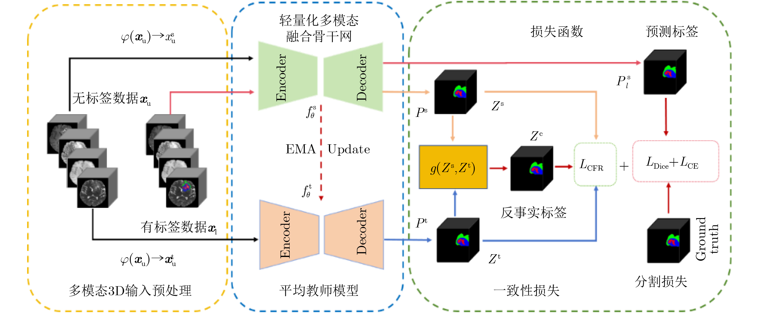
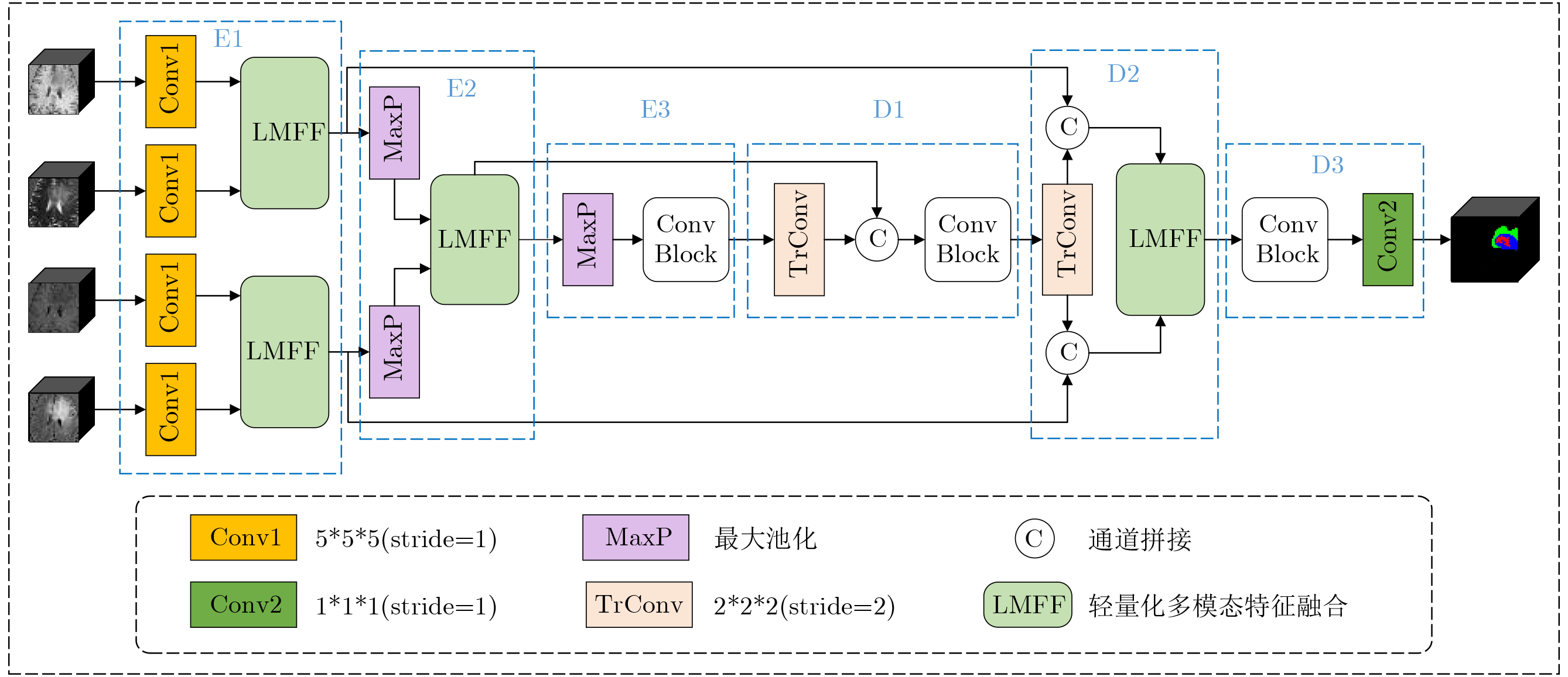
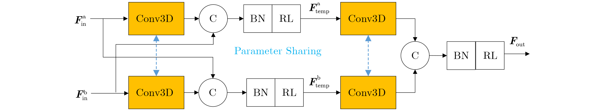
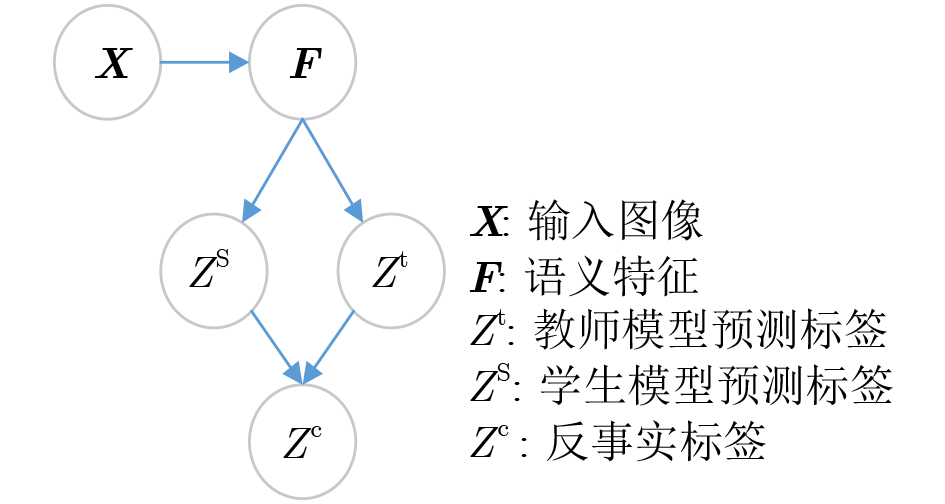
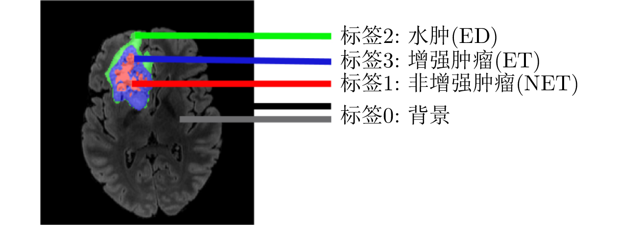
摘要: 针对脑肿瘤分割任务中标注样本稀缺、高计算开销以及病灶边界模糊问题,该文从模型结构与半监督机制两个角度出发,提出一种融合轻量化骨干网络与反事实推理的半监督脑肿瘤分割方法,旨在同时提升分割精度与模型部署效率。在网络结构设计方面,基于解剖结构一致性先验,构建了参数共享的多模态融合编码器-解码器架构,在保证分割性能的同时显著降低模型参数量与计算开销,使其适用于资源受限的临床应用场景。在半监督训练策略方面,利用教师-学生模型预测结果构建反事实样本,设计了一种结合像素级分割一致性与特征级语义稳定性的反事实推理损失函数,从而充分挖掘未标注数据的结构信息。在BraTS2021数据集上的实验结果表明,即使仅使用10%的标注数据,半监督模型在主要分割指标上平均达到约94%的全监督性能,同时在边界细节和小病灶识别性能方面均优于现有主流方法。Abstract:
Objective Brain tumor segmentation plays a key role in clinical diagnosis and treatment planning. However, reliable annotation of medical images is costly and time-consuming, which limits the availability of large annotated datasets. To address this problem, this paper proposes a semi-supervised brain tumor segmentation method that combines a lightweight multimodal fusion segmentation network with counterfactual reasoning. The aim is to improve segmentation accuracy while maintaining sufficient efficiency for deployment in resource-limited clinical scenarios. Methods A parameter-sharing multimodal encoder-decoder network is designed to reduce model size and computational cost. An anatomical-structure consistency prior is incorporated to improve alignment with brain anatomy. During training, a teacher-student framework is used to generate counterfactual samples from model predictions. These samples guide learning from unlabeled MRI scans through a counterfactual consistency loss that enforces pixel-level consistency and feature-level semantic stability. This strategy helps the model extract structural information from unlabeled data while reducing the risk of boundary distortion caused by conventional data augmentation. Results and Discussions Experiments on the BraTS 2019 and BraTS 2021 datasets show that the proposed method consistently outperforms comparison models under limited-label conditions. On BraTS 2019, the proposed method achieves the best average Dice Similarity Coefficient (DSC) of 66.06%, and its average Intersection over Union (IoU) of 53.16% is comparable to those of other models. More importantly, it obtains the lowest average 95% Hausdorff Distance (HD95) of 7.60 mm, representing reductions of approximately 11% and 6% compared with UNet3D and LightMUnet, respectively ( Tables 3 and4 ). On BraTS 2021, the semi-supervised model improves the average DSC and IoU by 4.51% and 5.29%, respectively, and reduces the average HD95 by 0.68 mm compared with the baseline model (Tables 5 and6 ). With only 10% labeled data, the proposed method achieves approximately 94% of the fully supervised performance in the main segmentation metrics. The model is also efficient, with only 1.657M parameters, a computational cost of 0.440 2 T, and an inference time of 0.093 7 s (Table 7 ). These results indicate that the proposed design achieves a favorable balance among segmentation accuracy, computational efficiency, and clinical deployment. The improvement is attributed to both the lightweight multimodal fusion segmentation network and the counterfactual mechanism, which guides the model to learn anatomically meaningful representations.Conclusions The proposed framework provides an effective solution for semi-supervised brain tumor segmentation. It balances accuracy, efficiency, and interpretability, and shows that causal reasoning can be integrated into medical image analysis in a practical manner. -
表 1 反事实标签
教师模型(Zt) 学生模型(Zs) 反事实标签(Zc) ET(3) ED(2) NET(1) ED(2) ET(3) NET(1) ED(2) NET(1) ET(3) NET(1) ED(2) ET(3) NET(1) ET(3) ED(2) ET(3) NET(1) ED(2)  下载: 导出CSV
表 2 数据集详情
数据集 样本总数 训练集 验证集 测试集 有标签 无标签 BraTS19 335 27 241 33 34 BraTS21 1251 100 900 125 126
下载: 导出CSV
表 3 BraTS 2019数据集上的结果对比(DSC和IoU指标对比)
模型 DSC(%)↑ IoU(%)↑ ET TC WT ET TC WT UNet3D(2015) 51.07±0.26 60.13±0.15 78.25±0.21 37.53±0.26 47.03±0.33 66.15±0.20 UXNet3D(2023) 58.86±0.19 61.55±0.08 77.15±0.15 46.14±0.25 48.46±0.06 65.32±0.77 LightMUnet(2024) 56.61±0.01 59.72±0.02 80.65±0.03 43.72±0.01 46.43±0.02 69.37±0.04 本文 55.40±0.18 62.64±0.13 80.13±0.05 41.68±0.14 49.50±0.07 68.32±0.13
下载: 导出CSV
表 4 BraTS 2019数据集上的结果对比(HD95和平均指标对比)
模型 HD95(mm)↓ 平均值 ET TC WT DSC(%)↑ IoU(%)↑ HD95(mm)↓ UNet3D(2015) 9.33±0.13 9.72±0.24 6.67±0.01 63.15±0.20 50.23±0.26 8.57±0.12 UXNet3D(2023) 12.12±0.21 11.86±0.31 8.95±0.10 65.85±0.14 53.30±0.56 10.97±0.20 LightMUnet(2024) 9.14±0.01 9.53±0.02 5.61±0.19 65.66±0.02 53.17±0.02 8.09±0.07 本文 9.67±0.10 8.33±0.17 4.79±0.14 66.06±0.12 53.16±0.11 7.60±0.13
下载: 导出CSV
表 5 BraTS 2021数据集上的结果对比(DSC和IoU)
模型 模式 有标签 无标签 DSC(%)↑ IoU(%)↑ ET TC WT ET TC WT UNet3D
(2015)base 100 0 64.54±0.20 68.29±0.11 82.05±0.13 54.67±0.21 55.25±0.14 71.62±0.18 semi 100 900 69.08±0.18 70.83±0.14 87.89±0.09 56.29±0.21 58.20±0.15 79.16±0.15 Attn-Unet
(2021)base 100 0 63.48±0.04 69.57±0.01 85.63±0.02 49.56±0.04 57.18±0.02 75.77±0.04 semi 100 900 68.84±0.34 69.79±0.08 86.10±0.07 55.88±0.52 57.52±0.10 76.36±0.17 UXNet3D
(2023)base 100 0 65.20±0.03 67.33±0.06 82.18±0.05 54.40±0.05 54.41±0.06 71.26±0.01 semi 100 900 68.88±0.05 74.85±0.04 86.95±0.06 60.15±0.06 62.95±0.04 77.82±0.09 LightMUnet
(2024)base 100 0 62.20±0.03 66.53±0.06 82.38±0.15 50.40±0.08 54.61±0.06 71.56±0.01 semi 100 900 67.88±0.07 73.75±0.09 85.65±0.06 54.15±0.06 62.25±0.04 76.52±0.21 本文 base 100 0 65.56±0.05 70.04±0.12 82.77±0.02 54.85±0.29 56.81±0.15 71.81±0.01 semi 100 900 69.30±0.16 77.30±0.16 88.28±0.05 60.46±0.19 66.05±0.26 79.70±0.08 full 1000 0 74.63±0.24 83.54±0.14 90.36±0.02 63.43±0.32 74.73±0.22 82.88±0.02
下载: 导出CSV
表 6 BraTS 2021数据集上的结果对比(HD95和平均指标)
模型 模式 有标签 无标签 HD95(mm)↓ 平均值 ET TC WT DSC(%)↑ IoU(%)↑ HD95(mm)↓ UNet3D
(2015)base 100 0 3.76±0.16 3.63±0.04 1.42±0.04 71.63±0.15 60.51±0.18 2.94±0.08 semi 100 900 2.85±0.04 2.81±0.02 1.09±0.01 75.93±0.14 64.55±0.17 2.25±0.02 Attn-Unet
(2021)base 100 0 4.46±0.01 4.89±0.01 3.82±0.01 72.89±0.02 60.84±0.03 4.46±0.01 semi 100 900 3.90±0.17 4.37±0.03 3.18±0.06 74.91±0.16 63.25±0.26 3.81±0.09 UXNet3D
(2023)base 100 0 3.70±0.04 3.51±0.24 1.71±0.04 71.57±0.05 60.02±0.04 2.97±0.11 semi 100 900 2.81±0.02 2.73±0.02 1.11±0.06 76.90±0.05 66.97±0.06 2.22±0.03 LightMUnet
(2024)base 100 0 4.07±0.04 4.01±0.13 2.17±0.14 70.37±0.08 58.86±0.05 3.42±0.10 semi 100 900 3.09±0.03 3.13±0.06 1.81±0.11 75.76±0.07 64.31±0.10 2.68±0.07 本文 base 100 0 3.42±0.03 3.51±0.01 1.05±0.01 72.79±0.06 61.16±0.15 2.66±0.02 semi 100 900 2.68±0.10 2.55±0.11 1.03±0.02 78.29±0.12 68.74±0.18 2.09±0.08 full 1000 0 2.08±0.05 2.13±0.01 1.00±0.01 82.84±0.13 73.68±0.19 1.74±0.02
下载: 导出CSV
表 7 性能指标对比结果
模型 参数量(M) 计算量(T) 推理时间 (s) UNet3D 6.5301 0.5643 4.0046 Attn-Unet 6.5920 0.5765 4.5678 UXNet3D 53.0594 1.3941 0.2780 LightM-Unet 6.1528 0.2343 3.3296 本文 1.6570 0.4402 0.0937
下载: 导出CSV
表 8 不同损失函数结果对比
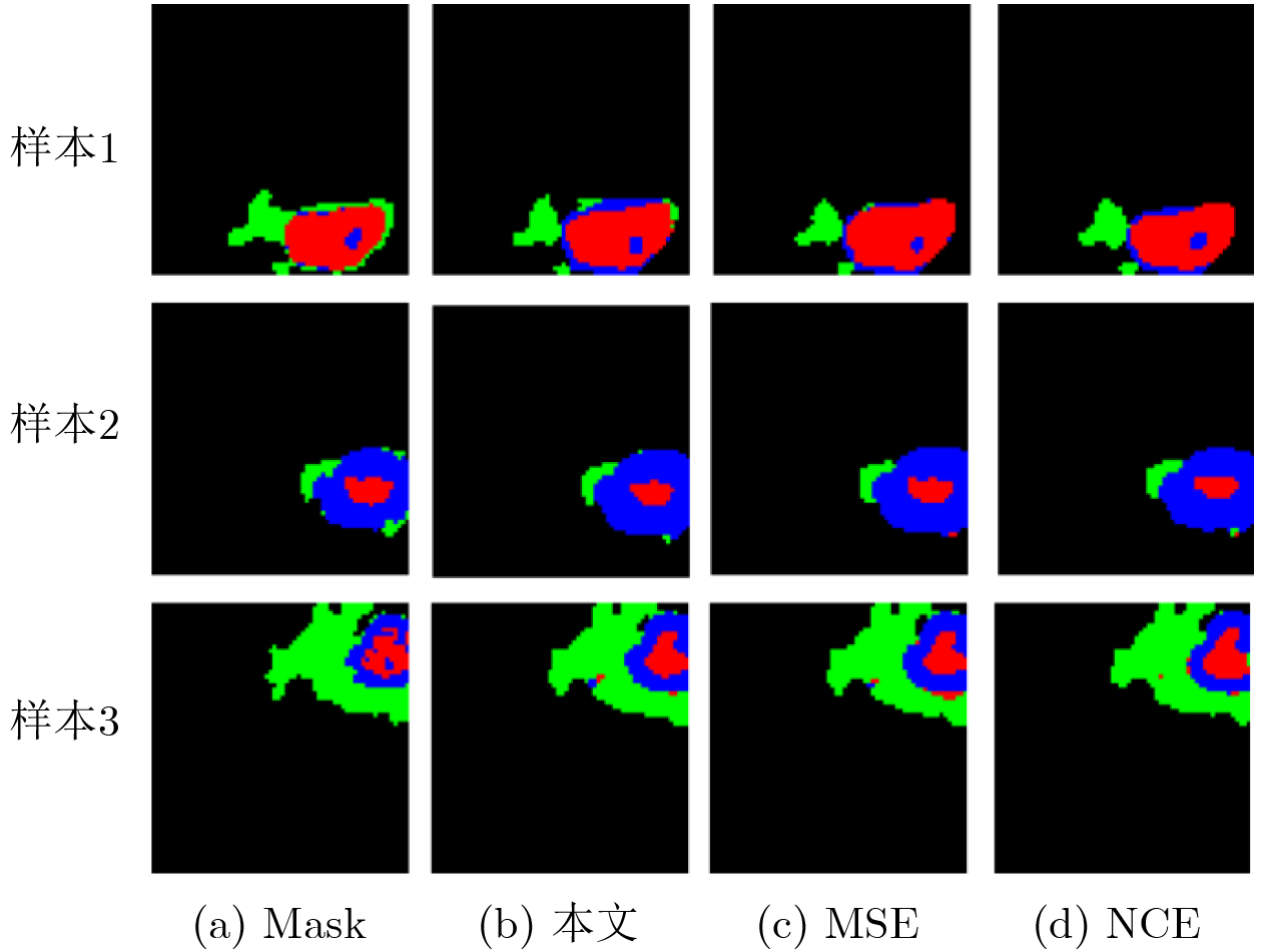
损失函数 分割区域 指标得分 DSC(%)↑ IOU(%)↑ HD95(mm)↓ MSE ET 69.07±0.24 56.09±0.29 2.74±0.09 TC 75.28±0.24 63.24±0.34 2.75±0.06 WT 87.65±0.01 78.72±0.04 1.05±0.03 NCE ET 69.21±0.18 56.23±0.33 2.73±0.12 TC 76.43±0.26 63.69±0.48 2.79±0.11 WT 87.31±0.09 78.32±0.07 1.06±0.08 本文 ET 69.30±0.16 56.46±0.19 2.68±0.10 TC 77.30±0.16 66.05±0.26 2.55±0.11 WT 88.28±0.05 79.70±0.08 1.03±0.02
下载: 导出CSV
-
[1] GHADIMI D J, VAHDANI A M, KARIMI H, et al. Deep learning-based techniques in glioma brain tumor segmentation using multi-parametric MRI: A review on clinical applications and future outlooks[J]. Journal of Magnetic Resonance Imaging, 2025, 61(3): 1094–1109. doi: 10.1002/JMRI.29543. [2] 江宗康, 吕晓钢, 张建新, 等. MRI脑肿瘤图像分割的深度学习方法综述[J]. 中国图象图形学报, 2020, 25(2): 215–228. doi: 10.11834/jig.190173.JIANG Zongkang, LV Xiaogang, ZHANG Jianxin, et al. Review of deep learning methods for MRI brain tumor image segmentation[J]. Journal of Image and Graphics, 2020, 25(2): 215–228. doi: 10.11834/jig.190173. [3] SOOMRO T A, ZHENG Lihong, AFIFI A J, et al. Image segmentation for MR brain tumor detection using machine learning: A review[J]. IEEE Reviews in Biomedical Engineering, 2023, 16: 70–90. doi: 10.1109/RBME.2022.3185292. [4] 张印辉, 张金凯, 何自芬, 等. 全局感知与稀疏特征关联图像级弱监督病理图像分割[J]. 电子与信息学报, 2024, 46(9): 3672–3682. doi: 10.11999/JEIT240364.ZHANG Yinhui, ZHANG Jinkai, HE Zifen, et al. Global perception and sparse feature associate image-level weakly supervised pathological image segmentation[J]. Journal of Electronics & Information Technology, 2024, 46(9): 3672–3682. doi: 10.11999/JEIT240364. [5] 丁建睿, 张听, 刘家栋, 等. 融合邻域注意力和状态空间模型的医学视频分割算法[J]. 电子与信息学报, 2025, 47(5): 1582–1595. doi: 10.11999/JEIT240755.DING Jianrui, ZHANG Ting, LIU Jiadong, et al. A medical video segmentation algorithm integrating neighborhood attention and state space model[J]. Journal of Electronics & Information Technology, 2025, 47(5): 1582–1595. doi: 10.11999/JEIT240755. [6] LEE H H, BAO Shunxing, HUO Yuankai, et al. 3D UX-Net: A large kernel volumetric convnet modernizing hierarchical transformer for medical image segmentation[C]. The 11th International Conference on Learning Representations, Kigali, Rwanda, 2023. [7] HATAMIZADEH A, TANG Yucheng, NATH V, et al. UNETR: Transformers for 3D medical image segmentation[C]. The IEEE/CVF Winter Conference on Applications of Computer Vision, Waikoloa, USA, 2022: 1748–1758. doi: 10.1109/WACV51458.2022.00181. [8] PUCH S, SÁNCHEZ I, HERNÁNDEZ A, et al. Global planar convolutions for improved context aggregation in brain tumor segmentation[C]. 4th International Workshop on Brainlesion: Glioma, Multiple Sclerosis, Stroke and Traumatic Brain Injuries, Granada, Spain, 2018: 393–405. doi: 10.1007/978-3-030-11726-9_35. [9] RONNEBERGER O, FISCHER P, and BROX T. U-Net: Convolutional networks for biomedical image segmentation[C]. 18th International Conference on Medical Image Computing and Computer-Assisted Intervention – MICCAI 2015, Munich, Germany, 2015: 234–241. doi: 10.1007/978-3-319-24574-4_28. [10] 刘海超, 宋丽娟. 多模态MRI脑肿瘤分割方法的特征融合技术综述[J]. 计算机工程与应用, 2024, 60(23): 28–48. doi: 10.3778/j.issn.1002-8331.2402-0087.LIU Haichao and SONG Lijuan. Review of feature fusion techniques for multimodal MRI brain tumor segmentation methods[J]. Computer Engineering and Applications, 2024, 60(23): 28–48. doi: 10.3778/j.issn.1002-8331.2402-0087. [11] LIU Yu, SHI Yu, MU Fuhao, et al. Multimodal MRI volumetric data fusion with convolutional neural networks[J]. IEEE Transactions on Instrumentation and Measurement, 2022, 71: 4006015. doi: 10.1109/TIM.2022.3184360. [12] 韩汶杞, 蒋雯, 耿杰, 等. 原型对齐与拓扑一致性约束下的多模态半监督遥感图像语义分割[J]. 电子与信息学报, 2025, 47(12): 4714–4727. doi: 10.11999/JEIT251115.HAN Wenqi, JIANG Wen, GENG Jie, et al. PATC: Prototype alignment and topology-consistent pseudo-supervision for multimodal semi-supervised semantic segmentation of remote sensing images[J]. Journal of Electronics & Information Technology, 2025, 47(12): 4714–4727. doi: 10.11999/JEIT251115. [13] ZHANG Zheng, YIN Guanchun, ZHANG Bo, et al. A semantic knowledge complementarity based decoupling framework for semi-supervised class-imbalanced medical image segmentation[C]. 2025 IEEE/CVF Conference on Computer Vision and Pattern Recognition (CVPR), Nashville, USA, 2025: 25940–25949. doi: 10.1109/CVPR52734.2025.02416. [14] TARVAINEN A and VALPOLA H. Mean teachers are better role models: Weight-averaged consistency targets improve semi-supervised deep learning results[C]. The 31st International Conference on Neural Information Processing Systems, Long Beach, USA, 2017: 1195–1204. [15] SOHN K, BERTHELOT D, LI Chunliang, et al. FixMatch: Simplifying semi-supervised learning with consistency and confidence[C]. The 34th International Conference on Neural Information Processing Systems, Vancouver, Canada, 2020: 51. [16] ASSEFA M, NASEER M, GANAPATHI I I, et al. DyCON: Dynamic uncertainty-aware consistency and contrastive learning for semi-supervised medical image segmentation[C]. 2025 IEEE/CVF Conference on Computer Vision and Pattern Recognition, Nashville, USA, 2025: 30850–30860. doi: 10.1109/CVPR52734.2025.02873. [17] CHI Hanyang, PANG Jian, ZHANG Bingfeng, et al. Adaptive bidirectional displacement for semi-supervised medical image segmentation[C]. 2024 IEEE/CVF Conference on Computer Vision and Pattern Recognition (CVPR), Seattle, USA, 2024: 4070–4080. doi: 10.1109/CVPR52733.2024.00390. [18] HITCHCOCK C and PEARL J. Causality: Models, reasoning and inference[J]. The Philosophical Review, 2001, 110(4): 639–641. doi: 10.2307/3182612. [19] JONES C, CASTRO D C, DE SOUSA RIBEIRO F, et al. A causal perspective on dataset bias in machine learning for medical imaging[J]. Nature Machine Intelligence, 2024, 6(2): 138–146. doi: 10.1038/s42256-024-00797-8. [20] OUYANG Cheng, CHEN Chen, LI Surui, et al. Causality-inspired single-source domain generalization for medical image segmentation[J]. IEEE Transactions on Medical Imaging, 2023, 42(4): 1095–1106. doi: 10.1109/TMI.2022.3224067. [21] MIAO Juzheng, CHEN Cheng, LIU Furui, et al. CauSSL: Causality-inspired semi-supervised learning for medical image segmentation[C]. The IEEE/CVF International Conference on Computer Vision, Paris, France, 2023: 21369–21380. doi: 10.1109/ICCV51070.2023.01959. [22] QU Jiaqi, XIAO Xiang, WEI Xunbin, et al. A causality-inspired generalized model for automated pancreatic cancer diagnosis[J]. Medical Image Analysis, 2024, 94: 103154. doi: 10.1016/j.media.2024.103154. [23] WANG Sihan, LI Lei, and ZHUANG Xiahai. AttU-NET: Attention U-Net for brain tumor segmentation[C]. 7th International Workshop on Brainlesion: Glioma, Multiple Sclerosis, Stroke and Traumatic Brain Injuries, 2021: 302–311. doi: 10.1007/978-3-031-09002-8_27. [24] LIAO Weibin, ZHU Yinghao, WANG Xinyuan, et al. LightM-UNet: Mamba assists in lightweight UNet for medical image segmentation[J]. arXiv preprint arXiv: 2403.05246, 2024. doi: 10.48550/arXiv.2403.05246. -

 下载:
下载:


图(10) / 表(8)
计量
- 文章访问数: 789
- HTML全文浏览量: 273
- PDF下载量: 74
- 被引次数: 0
