

Research on Ultrasound Imaging Algorithm Fused with Diffusion Model
-
摘要: 针对超声成像分辨率低及易受伪影干扰问题,该文提出基于扩散模型(DM)的U-DM超声成像质量优化方法。通过构建差值训练机制与解剖结构引导策略,结合改进型U-Net网络架构实现多尺度特征融合,建立从含噪超声数据到高质量图像的映射关系,进而生成高质量超声图像。基于PICMUS数据集的实验结果表明,该文所提U-DM方法在噪声抑制与结构保持方面显著优于UNet和UNet-GAN等方法,能有效消除人工伪影并恢复解剖细节,其图像重建质量达到临床诊断要求。相较于生成对抗网络(GAN),该文所提融合扩散模型的超声成像方法展现出更稳定的训练特性和更优的泛化能力,克服了模式坍塌等固有问题,为突破超声成像质量瓶颈提供了新途径。Abstract:
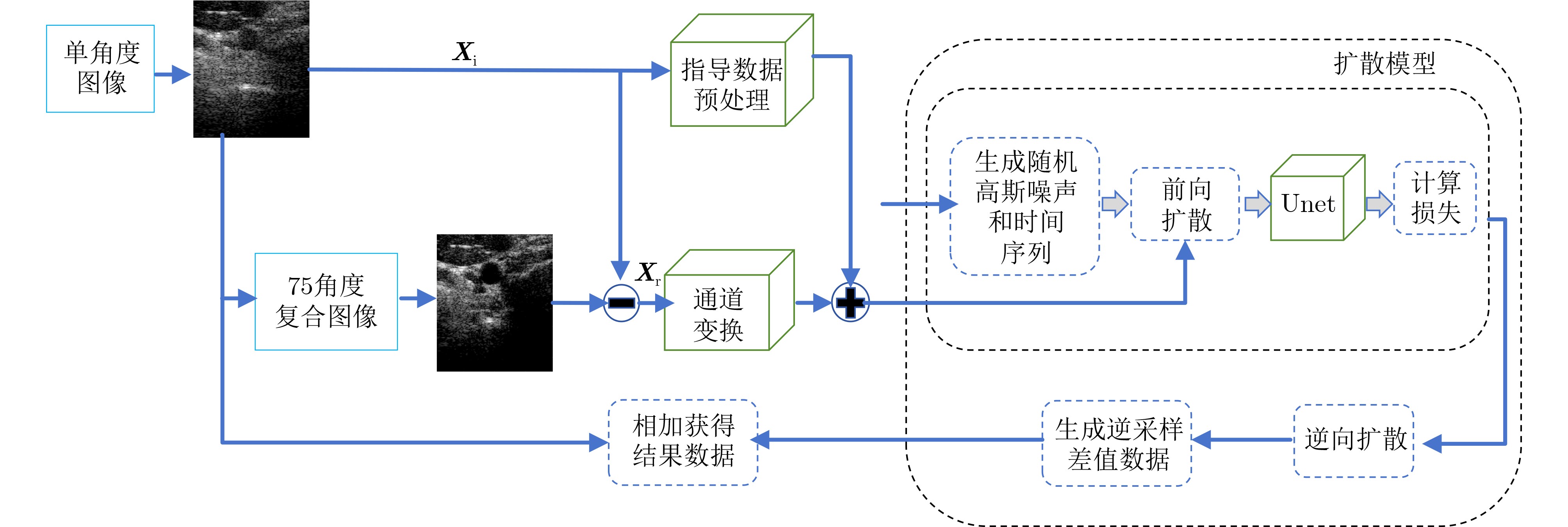
Objective Medical ultrasound imaging uses ultrasonic waves to probe human tissues and forms images by processing returning echoes. It has become an essential clinical diagnostic tool because it is noninvasive, safe, and capable of real-time imaging. However, conventional ultrasound imaging remains fundamentally limited by factors such as the finite width of ultrasonic pulses, variations in tissue acoustic impedance, and the complexity of echo signals. These factors lead to persistent challenges, including limited spatial resolution, severe speckle noise, and off-axis artifacts. These limitations directly reduce lesion detectability and diagnostic accuracy. Traditional approaches based on hardware optimization and signal processing algorithms, such as adaptive beamforming, have provided only incremental improvement. Their performance is often constrained by physical laws, computational complexity, and dependence on manual parameter tuning. Recent deep learning methods, particularly those based on Generative Adversarial Networks (GANs), have shown promising performance, but they suffer from training instability and limited interpretability. The diffusion model, an emerging state-of-the-art generative framework, has shown strong robustness and generalization in Computed Tomography (CT) and Magnetic Resonance Imaging (MRI) reconstruction. However, its application in ultrasound imaging remains largely unexplored. This study aims to address this gap by developing a novel diffusion model-based framework for high-quality ultrasound image formation and to provide a stable, efficient, and interpretable solution for improving ultrasound image quality. Methods A novel ultrasound imaging method based on a Denoising Diffusion Probabilistic Model (DDPM) is proposed. The core of the method is a multi-scale diffusion network architecture designed to progressively refine a low-quality ultrasound image, such as one generated by a simple Delay-And-Sum (DAS) beamformer, into a high-quality image. The process includes forward and reverse stages. In the forward stage, Gaussian noise is gradually added to a high-quality ground-truth image over a series of time steps. In the reverse stage, the model is trained to learn the conditional denoising function. A custom denoising network takes a low-resolution DAS image as conditional input and fuses it with the noisy image at each denoising step through residual connections and feature-wise transformations at multiple scales. This deep fusion mechanism enables the network to incorporate the underlying anatomical structure from the low-quality input while iteratively removing noise and artifacts through the diffusion process. The model is trained on a dataset of paired low-quality and high-quality ultrasound images, in which the high-quality images serve as the training target. The training objective is to maximize the variational lower bound of the likelihood, thereby enabling the network to reverse the noising process. The proposed method is quantitatively compared with traditional DAS, Minimum Variance (MV) beamforming, and a representative GAN-based super-resolution method using Peak Signal-to-Noise Ratio (PSNR) and Structural SIMilarity index (SSIM). Results and Discussions The proposed diffusion model demonstrates superior performance in improving ultrasound image quality. Quantitatively, the method achieves a mean PSNR of 35.2 dB and an SSIM of 0.933, with a PSNR improvement of 4.5 dB over conventional beamforming methods, while maintaining excellent structural fidelity. The method also consistently outperforms adaptive MV beamforming and GAN-based methods across all evaluation metrics, including contrast-to-noise ratio. Visual assessment supports these quantitative results. The generated images show markedly reduced speckle noise and substantially improved boundary definition of anatomical structures. Notably, these improvements are achieved without the blurring or artificial textures commonly observed in other deep learning-based methods. The multi-scale architecture with conditional feature injection effectively preserves structural integrity, as shown by the clear and continuous edges in the output images. The progressive denoising nature of the method also provides inherent interpretability for the image refinement process. Unlike the opaque single-step generation used in many other deep learning models, this method provides a transparent, stepwise enhancement pathway from the initial input to the final output. In addition, the training process remains stable and convergent, avoiding the instability that frequently affects adversarial training methods. Ablation experiments confirm the critical role of the deep fusion mechanism, and resolution analysis verifies substantial improvement in both lateral and axial resolution compared with all baseline methods. Conclusions This study develops and validates a novel ultrasound imaging method based on a diffusion model. The proposed framework effectively addresses key limitations of conventional methods and existing deep learning-based approaches. It avoids the complex matrix computations and manual parameter tuning required by adaptive beamformers and provides a more stable training framework than GAN-based methods. The results show that the method can substantially improve image quality by increasing PSNR and maintaining excellent structural similarity, thereby producing images with suppressed noise, reduced artifacts, and improved resolution. The multi-scale diffusion process preserves anatomical structures and provides a degree of interpretability for the image generation process. This work establishes diffusion models as a promising new framework for advanced ultrasound imaging and provides a robust, high-performance technical route for overcoming current bottlenecks in ultrasound image quality, with broad potential clinical value. -
Key words:
- Ultrasound imaging /
- Diffusion Model (DM) /
- UNet /
- Difference training mechanism
-
表 1 基于颈动脉数据的PSNR和SSIM指标对比结果
指标 1 PW U-DM 颈动脉横截面PSNR(dB) 21.343 35.423 颈动脉纵截面PSNR(dB) 19.884 34.944 颈动脉横截面SSIM 0.730 0.931 颈动脉纵截面SSIM 0.709 0.927  下载: 导出CSV
表 2 囊肿体膜数据的CR和CNR指标结果
指标 1 PW U-DM 真实囊肿体膜CR(dB) 14.293 30.849 模拟囊肿体膜CR(dB) 16.823 36.502 真实囊肿体膜CNR 1.797 3.533 模拟囊肿体膜CNR 1.093 1.679
下载: 导出CSV
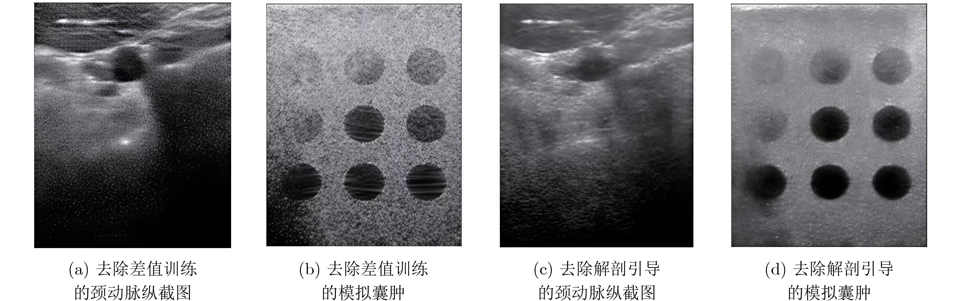
表 3 消融实验结果
模型配置 PSNR(dB) SSIM CR(dB) CNR 完整U-DM模型 34.948 0.927 27.032 3.335 去除差值训练 26.295 0.773 24.468 2.646 去除解剖引导 23.469 0.675 22.259 2.523
下载: 导出CSV
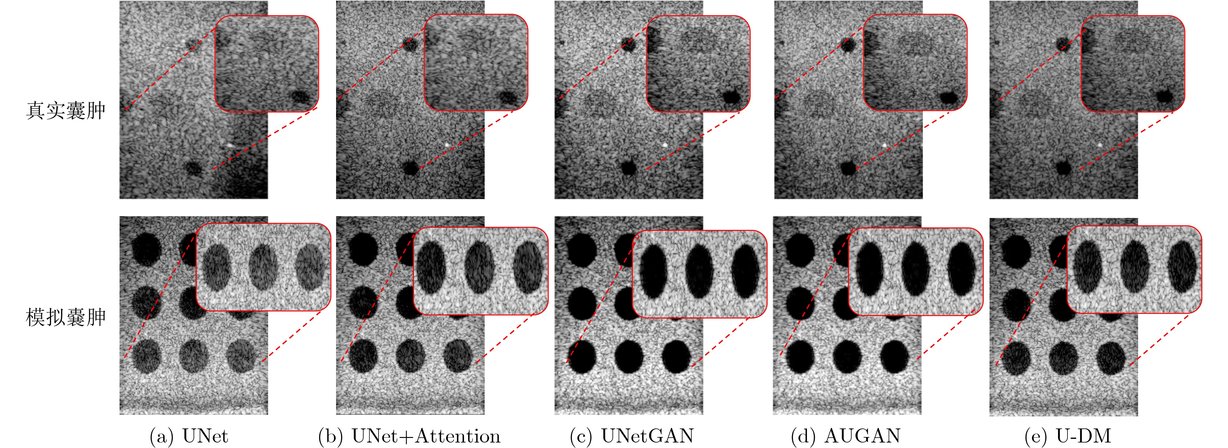
表 4 U-DM方法与其他网络结构在颈动脉图像的PSNR, SSIM指标上的对比
指标 UNet UNet+Attention UNetGAN AUGAN U-DM 颈动脉横截面PSNR(dB) 30.514 32.239 33.943 34.128 35.423 颈动脉纵截面PSNR(dB) 29.668 31.125 33.509 33.486 34.944 颈动脉横截面SSIM 0.872 0.888 0.912 0.921 0.931 颈动脉纵截面SSIM 0.861 0.873 0.903 0.905 0.927
下载: 导出CSV
表 5 囊肿图像的CR, CNR指标对比
指标 UNet UNet+Attention UNetGAN AUGAN U-DM 真实囊肿体膜
CR(dB)28.820 30.445 31.061 31.496 30.849 模拟囊肿体膜
CR(dB)35.318 36.034 44.507 44.873 36.502 真实囊肿体膜
CNR2.721 3.110 3.429 3.481 3.533 模拟囊肿体膜
CNR1.771 1.798 1.992 2.087 1.897
下载: 导出CSV
表 6 不同模型在PICMUS各场景下的性能变异系数CV(%)
方法 PSNR(dB) SSIM CR(dB) CNR 综合 AUGAN 11.0 5.3 10.1 17.0 10.9 UNetGAN 10.6 4.0 8.2 18.6 10.4 UNet+Attention 2.6 1.4 6.0 5.5 3.9 U-DM 3.8 1.3 7.1 2.3 3.6 UNet 0.7 0.2 6.1 5.8 3.2
下载: 导出CSV
-
[1] 刘佳敏, 吴佩先, 曾凡勇. 多模态超声成像在甲状腺结节良恶性鉴别诊断中的研究进展[J]. 影像研究与医学应用, 2025, 9(24): 8-10. doi: 10.20267/j.issn.2096-3807.2025.24.003.LIU Jiamin, WU Peixian, and ZENG Fanyong. Research progress of multimodal ultrasound in the differential diagnosis of benign and malignant thyroid nodules[J]. Journal of Imaging Research and Medical Applications, 2025, 49(24): 8-10. doi: 10.20267/j.issn.2096-3807.2025.24.003. [2] Zhang, Jingke, He, Qiong, Xiao, Yang, et al. Ultrasound image reconstruction from plane wave radio-frequency data by self-supervised deep neural network[J]. Medical image analysis, 2021, 70. doi: 10.1016/j.media.2021.102018. [3] 张豪洁, 周箩鱼. 超声测井图像过井裂缝提取算法研究[J]. 光电子.激光, 2019, 30(6): 654-658. doi: 10.16136/j.joel.2019.06.0349.ZHANG Haojie and ZHOU Luoyu. Research on fracture extraction algorithm from ultrasonic logging images[J]. Journal of Optoelectronics·Laser, 2019, 30(6): 654-658. doi: 10.16136/j.joel.2019.06.0349. [4] Chu, Xuan, Wang, Tengfei, Chen, Meiwen, et al. Deep learning model for malignancy prediction of TI-RADS 4 thyroid nodules with high-risk characteristics using multimodal ultrasound: A multicentre study[J]. Computerized Medical Imaging and Graphics: The Official Jounal of the Computerized Medical Imaging Society, 2025, 124102576. doi: 10.1016/j.compmedimag.2025.102576. [5] 陈尧, 熊政辉, 罗俊威, 等. 复杂型面航空构件自动化超声成像检测技术研究进展[J]. 航空材料学报, 2025, 45(6): 33-44. doi: 10.11868/j.issn.1005-5053.2024.000173.CHEN Yao, XIONG Zhenghui, LUO Junwei, et al. Research progress on automated ultrasonic imaging detection technology for complex-shaped aerospace components[J]. Journal of Aeronautical Materials, 2025, 45(6): 33-44. doi: 10.11868/j.issn.1005-5053.2024.000173. [6] MORA P, CHEKROUN M, RAETZ S, et al. Nonlinear generation of a zero group velocity mode in an elastic plate by non-collinear mixing[J]. Ultrasonics, 2022, 119: 106589. doi: 10.1016/j.ultras.2021.106589. [7] 张克潜, 李德杰. 微波与光电子学中的电磁理论[M]. 2版. 北京: 电子工业出版社, 2001: 210–215.ZHANG Keqian and LI Dejie. Electromagnetic Theory for Microwaves and Optoelectronics[M]. 2nd ed. Beijing: Publishing House of Electronics Industry, 2001: 210–215. [8] Liu R L , Wu Y Q , Liu J H , et al. The segmentation of FMI image based on 2-D dyadic wavelet transform[J]. Applied Geophysics, 2005, 2(2): 89-93. doi: 10.1007/s11770-005-0039-z. [9] 王冬冬, 刘世伟, 侯振永. 多功能超声成像测井仪在塔里木油田应用效果评价[J]. 测井技术, 2023, 47(3): 364–370 doi: 10.16489/j.issn.1004-1338.2023.03.016.WANG Dongdong, LIU Shiwei, and HOU Zhenyong. Application effect evaluation of multifunctional ultrasonic imaging logging tool in Tarim Oilfield[J]. Well Logging Technology, 2023, 47(3): 364–370. doi: 10.16489/j.issn.1004-1338.2023.03.016. [10] Ben Luijten, Regev Cohen, Frederik J. de Bruijn, et al. Adaptive Ultrasound Beamforming Using Deep Learning[J]. IEEE Transactions on Medical Imaging, 2020, 39(12): 3967-3978. doi: 10.1109/TMI.2020.3008537. [11] HO D J, MONTSERRAT D M, FU Chichen, et al. Sphere estimation network: Three-dimensional nuclei detection of fluorescence microscopy images[J]. Journal of Medical Imaging, 2020, 7(4): 044003. doi: 10.1117/1.JMI.7.4.044003. [12] Camacho J , Cruza J F , Brizuela J , et al.Automatic Dynamic Depth Focusing for NDT[J].IEEE Transactions on Ultrasonics Ferroelectrics and Frequency Control, 2014, 61(4): 673-684. doi: 10.1109/TUFFC.2014.2955. [13] 曹欢庆, 朱启民, 赵培含, 等. 复杂型面结构超声成像检测研究进展[J]. 仪器仪表学报, 2024, 45(6): 42-53. doi: 10.19650/j.cnki.cjsi.J2412566.CAO Huanqing, ZHU Qimin, ZHAO Peihan, et al. Research progress on ultrasonic imaging detection of complex surface structures[J]. Chinese Journal of Scientific Instrument, 2024, 45(6): 42-53. doi: 10.19650/j.cnki.cjsi.J2412566. [14] Ronneberger O , Fischer P , Brox T .U-Net: Convolutional Networks for Biomedical Image Segmentation[J].Springer, Cham, 2015. doi: 10.1007/978-3-662-54345-0_3. [15] BILLOT B, GREVE D N, PUONTI O, et al. SynthSeg: Segmentation of brain MRI scans of any contrast and resolution without retraining[J]. Medical Image Analysis, 2023, 86: 102789. doi: 10.1016/j.media.2023.102789. [16] van Sloun R J G, Cohen R, Eldar Y C. Deep learning in ultrasound imaging [J]. Proceedings of the IEEE, 2020, 108 (1): 11-29 [17] 衡佳鸣, 王宁浩, 董凤林, 等. 基于深度学习的超声成像技术研究现状 [J]. 声学技术, 2023, 42 (2): 174-183. doi: 10.16300/j.cnki.1000-3630.2023.02.008.HENG Jiaming, WANG Ninghao, DONG Fenglin, et al. Research status of ultrasound imaging technology based on deep learning[J]. Technical Acoustics, 2023, 42(2): 174-183. doi: 10.16300/j.cnki.1000-3630.2023.02.008. [18] 段鹤立, 牛逸凡, 王瑞琦, 等. 多模态超声成像技术鉴别Graves病与慢性淋巴细胞性甲状腺炎的研究进展[J]. 中国医疗设备, 2024, 39(5): 175-180. doi: 10.3969/j.issn.1674-1633.2024.05.029.DUAN Heli, NIU Yifan, WANG Ruiqi, et al. Research progress of multimodal ultrasound imaging in differentiating Graves’ disease and chronic lymphocytic thyroiditis[J]. China Medical Devices, 2024, 39(5): 175-180. doi: 10.3969/j.issn.1674-1633.2024.05.029. [19] 武林松, 王冬, 彭艳艳, 等. SWE联合SMI在甲状腺良恶性结节鉴别诊断中的应用[J]. 中国医科大学学报, 2024, 53(6): 541-546. doi: 10.12007/j.issn.0258-4646.2024.06.010.WU Linsong, WANG Dong, PENG Yanyan, et al. Application of SWE combined with SMI in the differential diagnosis of benign and malignant thyroid nodules[J]. Journal of China Medical University, 2024, 53(6): 541-546. doi: 10.12007/j.issn.0258-4646.2024.06.010. [20] Cruz R A Q , Cacau D C , Santos R M D , et al.Improving Accuracy of Automatic Fracture Detection in Borehole Images with Deep Learning and GPUs[J].IEEE Computer Society, 2017. doi: 10.1109/sibgrapi.2017.52. [21] Goodfellow, Ian, Pouget-Abadie, Jean, Mirza, Mehdi, et al. Generative Adversarial Networks[J]. Communications of the ACM, 2020, 63(11): 139-144. doi: 10.1145/3422622. [22] 宋佳好, 任芸芸. 胎盘-心脏轴及胎盘超声成像技术的研究进展[J]. 复旦学报(医学版), 2024, 51(5): 825-830. doi: 10.3969/j.issn.1672-8467.2024.05.027.SONG Jiahao and REN Yunyun. Research progress on the placental-heart axis and placental ultrasound imaging technology[J]. Fudan Journal (Medical Sciences), 2024, 51(5): 825-830. doi: 10.3969/j.issn.1672-8467.2024.05.027. [23] Hyun, Dongwoon, Brickson, Leandra L. , Looby, Kevin T., et al. Beamforming and Speckle Reduction Using Neural Networks[J]. IEEE Transactions on Ultrasonics, Ferroelectrics, and Frequency Control, 2019, 66(5): 898–910. doi: 10.1109/TUFFC.2019.2903795. [24] 张经科, 何琼, 罗建文. 平面波超声成像中的波束合成方法研究进展[J]. 应用声学, 2021, 40(1): 22-32. doi: 10.11684/j.issn.1000-310X.2021.01.003.ZHANG Jingke, HE Qiong, and LUO Jianwen. Research progress on beamforming methods in plane wave ultrasound imaging[J]. Journal of Applied Acoustics, 2021, 40(1): 22-32. doi: 10.11684/j.issn.1000-310X.2021.01.003. [25] 王希吉. 超声成像测井技术在地质勘探中的应用[J]. 煤炭经济研究, 2024, 44(z1): 155-161. doi: 10.3969/j.issn.1002-9605.2024.z1.032.WANG Xiji. Application of ultrasonic imaging logging technology in geological exploration[J]. Coal Economic Research, 2024, 44(z1): 155-161. doi: 10.3969/j.issn.1002-9605.2024.z1.032. [26] 杨磊, 宋昊, 申瑞阳, 等. 强稀疏低副瓣近场聚焦稀疏阵列三维成像[J]. 电子与信息学报, 2024, 46(12): 4471–4482. doi: 10.11999/JEIT231278.YANG Lei, SONG Hao, SHEN Ruiyang, et al. High sparsity and low sidelobe near-field focused sparse array for three-dimensional imagery[J]. Journal of Electronics & Information Technology, 2024, 46(12): 4471–4482. doi: 10.11999/JEIT231278. -

 下载:
下载:


图(9) / 表(6)
计量
- 文章访问数: 491
- HTML全文浏览量: 250
- PDF下载量: 80
- 被引次数: 0
