

A Causality-Guided KAN Attention Framework for Brain Tumor Classification
-
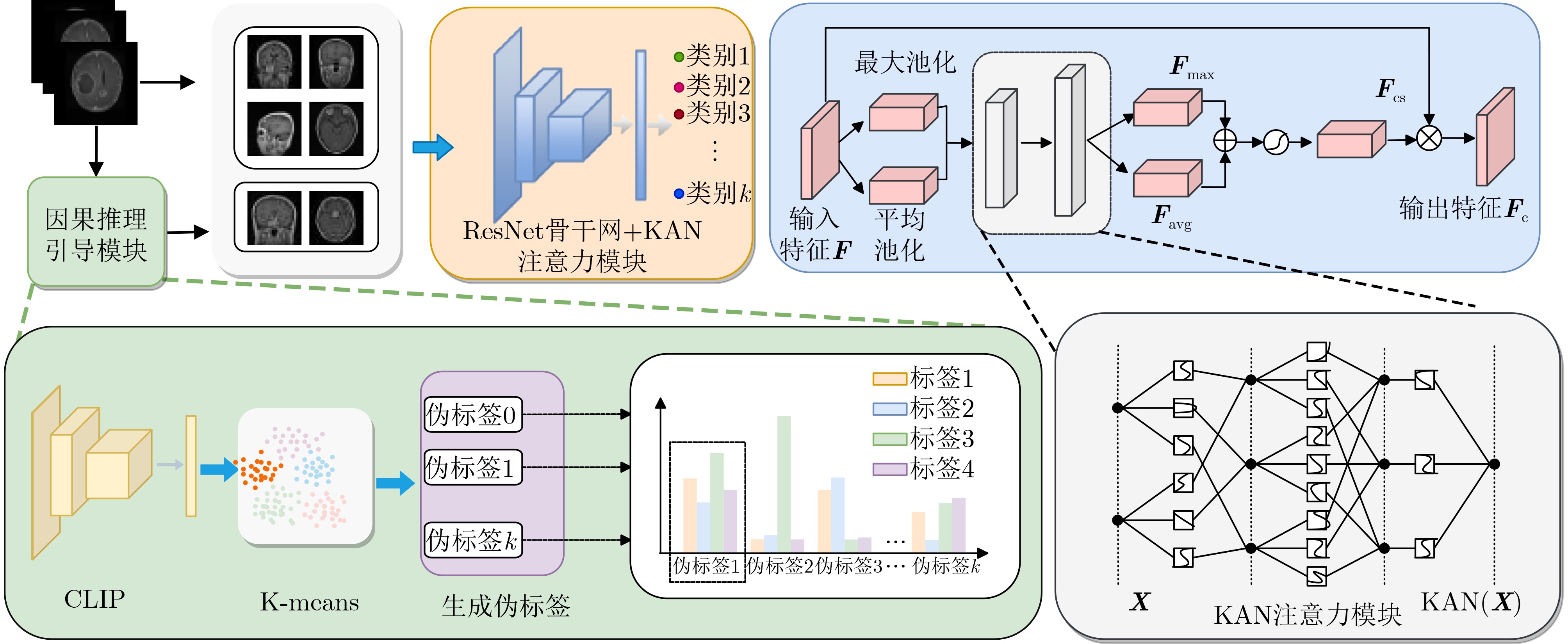
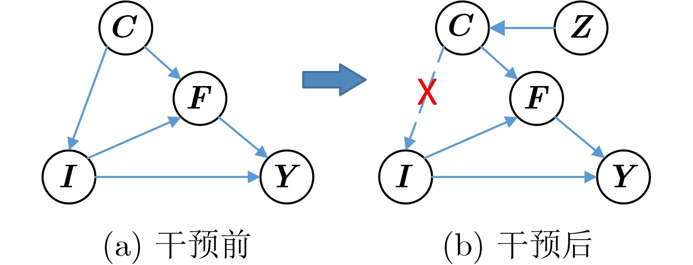
摘要: 脑肿瘤分类是医学影像分析中的关键任务,但现有深度学习方法在应对扫描参数差异、解剖位置偏移等因素时仍面临特征混淆问题,且难以建模肿瘤异质性引发的复杂非线性关系。针对这一挑战,该文提出一种因果推理引导的KAN注意力分类框架。首先,基于CLIP模型进行无监督特征提取,捕捉MRI数据中的高层语义特征;其次,基于K-means聚类设计混淆均衡度指标,筛选混淆因子图像,并设计因果干预机制,显式引入混淆样本,同时提出因果增强的损失函数以优化模型的判别能力;最后,在预训练ResNet主干网中引入KAN注意力模块,强化模型对肿瘤局部坏死区与强化边缘的非线性关联建模能力。实验表明,所提出的方法在脑肿瘤分类任务中优于传统CNN与Transformer模型,验证了其在判别能力和鲁棒性方面的优势。该研究为医学影像的因果推理与高阶非线性建模提供了新的技术路径。Abstract:
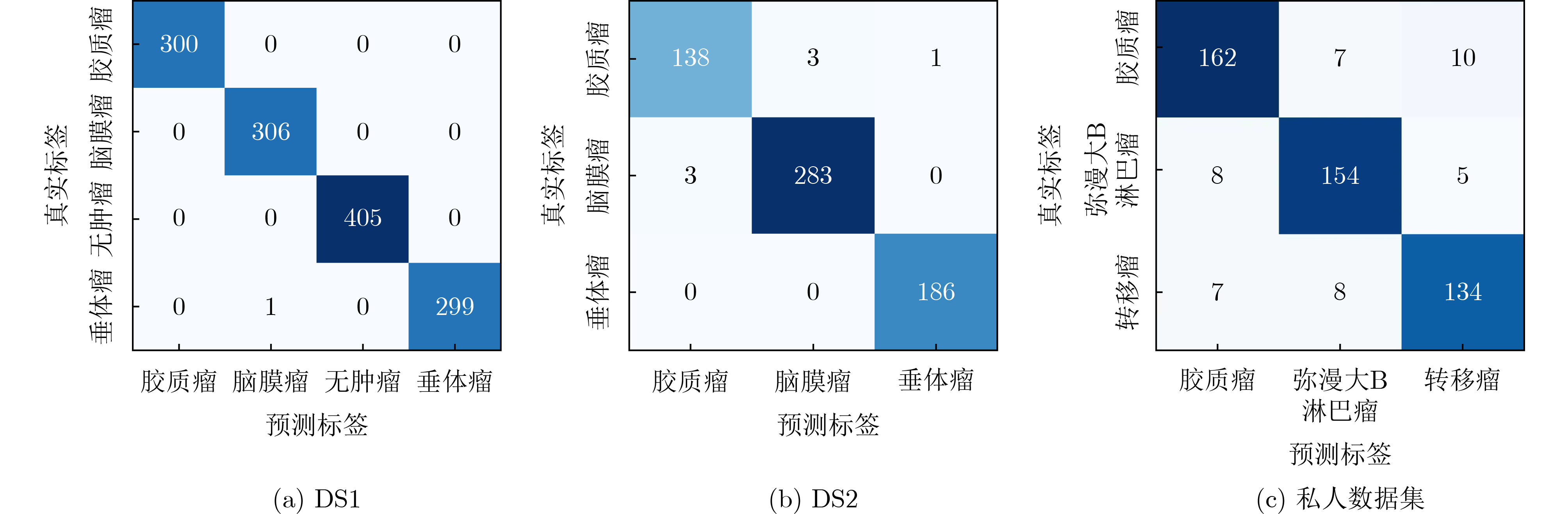
Objective Convolutional Neural Network (CNN)-based Computer-Aided Diagnosis (CAD) systems have advanced brain tumor classification in recent years. However, performance remains limited by feature confusion and insufficient modeling of high-order interactions. This study proposes a framework that integrates causal feature guidance with a KAN attention mechanism. A Confusion Balance Index (CBI) is developed to quantify real label distribution within clusters. A causal intervention mechanism then incorporates confused samples to strengthen discrimination between causal variables and confounding factors. A spline-based KAN attention module is further constructed to model high-order feature interactions and enhance focus on critical lesion regions and discriminative features. The combined causal modeling and nonlinear interaction enhancement improves robustness and addresses the inability of traditional architectures to capture complex pathological feature relationships. Methods A pre-trained CLIP model is used for feature extraction to obtain semantically rich visual representations. K-means clustering and the CBI are applied to identify confusing factor images, after which a causal intervention mechanism incorporates these samples into the training process. A causal-enhanced loss function is then designed to strengthen discrimination between causal variables and confounding factors. To address limited high-order interaction modeling, a Kolmogorov-Arnold Network (KAN)-based attention mechanism is integrated. This spline-based module constructs flexible nonlinear attention representations and refines high-order feature interactions. When fused with the backbone network, it improves discriminative performance and generalization. Results and Discussions The proposed method achieves superior performance across three datasets. On DS1, the model reaches 99.92% accuracy, 99.98% specificity, and 99.92% precision, outperforming RanMerFormer (+0.15%) and SAlexNet (+0.23%) and exceeding traditional CNNs by more than 2% (95%~97%). Swin Transformers reach 98.08% accuracy but only 91.75% precision, indicating stronger robustness of the proposed model in reducing false detections. On DS2, the method achieves 98.86% accuracy and 98.80% precision, exceeding the next-best RanMerFormer. On a more challenging in-house dataset, it maintains 90.91% accuracy and 95.45% specificity, showing generalization in complex settings. The gains result from the KAN attention mechanism’s ability to model high-order interactions and the causal reasoning module’s decoupling of confounding factors. These components improve focus on lesion regions and stabilize decision-making in complex scenarios. The results demonstrate reliable performance for clinical precision diagnostics. Conclusions The findings confirm that the proposed framework improves brain tumor classification. The combined effect of the causal intervention mechanism and the KAN attention module is the primary contributor to performance gains. These improvements require minimal increases in model parameters and inference latency, preserving efficiency and practicality. The study proposes a methodological direction for medical image classification and shows potential utility in few-shot learning and clinical decision support. -
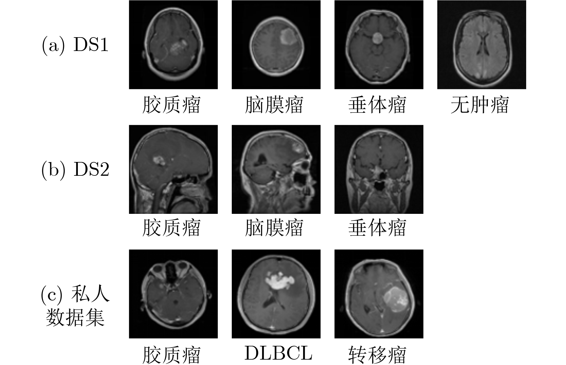
表 1 数据集信息
肿瘤类型 DS1 DS2 私人 脑膜瘤 1645 708 - 垂体瘤 1757 930 - 胶质瘤 1621 1426 891 DLBCL - - 468 转移瘤 - - 743 无肿瘤 2000 - - 总 数 7023 3064 2102 训练集 5712 2451 1680 测试集 1311 613 422  下载: 导出CSV
表 2 不同模型的测试集结果比较(%)
数据集 方法 Pr Se Sp Acc DS1 FTVT[24] 98.71 98.70 - 98.70 Swin Transformer V2[25] 98.75 98.51 - 98.97 NeuroNet19[26] 99.20 99.20 - 99.30 ResVit[27] 98.45 98.61 - 98.47 InceptionV3[28] 97.97 96.59 99.98 97.13 CNN[29] 97.00 97.00 - 97.00 DCST+SVM [30] 97.80 96.62 - 97.71 Swin Transformers [12] 91.75 - - 98.08 Custom built CNN[31] 95.00 95.00 - 95.16 RanMerFormer [14] 99.76 99.75 99.93 99.77 SAlexNet[9] 99.37 99.33 - 99.69 TinyViT*[32] 99.43 99.44 99.83 99.47 LeViT*[33] 98.92 98.93 99.68 99.01 Mobilenet-v4*[34] 97.99 97.98 99.37 98.09 本文方法 99.92 99.92 99.98 99.92 DS2 ARM-Net[35] 96.46 96.09 - 96.64 GT-Net[36] - - - 97.11 ResVit[27] 98.54 98.54 - 98.53 GAN+ConvNet[37] 95.29 94.91 97.69 95.60 CNN+SVM[38] 97.30 97.60 98.97 98.00 RanMerFormer[14] 98.87 98.46 99.39 98.86 Gaussian CNN[39] 97.07 - - 97.82 DACBT[40] - 98.09 100 98.56 Custom built CNN[10] 96.06 94.43 96.93 96.13 VGG19+CNN[41] 98.34 98.60 99.28 98.54 GoogleNet[42] 97.20 97.30 98.96 97.10 TinyViT*[32] 97.73 98.01 99.02 98.05 LeViT*[33] 97.10 97.07 98.68 97.39 Mobilenet-v4*[34] 97.45 97.19 98.70 97.56 本文方法 98.80 98.70 99.40 98.86 私人数据集 VIT*[43] 80.88 80.85 90.60 81.21 GoogleNet*[42] 90.09 90.29 95.10 90.10 TinyViT*[28] 88.73 88.33 94.20 88.48 LeViT*[29] 80.89 80.94 90.57 81.01 Mobilenet-v4*[34] 86.72 86.72 93.35 86.46 本文方法 90.86 90.88 95.45 90.91 注:“*”表示在统一实验设置下复现得到的结果。粗体代表性能最优值。
下载: 导出CSV
表 3 消融实验结果比较(%)
数据集 方法 Pr Se Sp Acc DS1 ResNet18 99.84 99.83 99.95 99.85 ResNet18+Causal 99.92 99.92 99.98 99.92 ResNet18+KAM 99.92 99.92 99.98 99.92 ResNet18+Causal+KAM 99.92 99.92 99.98 99.92 DS2 ResNet18 98.29 98.13 99.15 98.37 ResNet18+Causal 98.37 98.42 99.24 98.53 ResNet18+KAM 98.41 98.48 99.23 98.53 ResNet18+Causal+KAM 98.76 98.71 99.41 98.86 私人数据集 ResNet18 87.28 87.46 93.66 87.27 ResNet18+Causal 89.58 89.72 94.89 89.70 ResNet18+KAM 89.61 89.79 94.88 89.70 ResNet18+Causal+KAM 90.86 90.88 95.45 90.91
下载: 导出CSV
表 4 模型性能分析
模型 平均Acc(%) 参数量(M) FLOPs(G) ResNet18 95.15 11.178 1.8186 ResNet18+CAM 96.02 11.222 1.8190 ResNet+SE 92.05 11.222 1.8240 ResNet+CBAM 95.93 11.267 1.8240 ResNet18+KAM 96.56 11.188 1.8189 注:加粗数值表示最优值。
下载: 导出CSV
表 5 不同因果权重$ \alpha $下的分类准确率
超参数α Acc (%) 超参数$ \alpha $ 0.0 0.1 0.3 0.5 0.7 0.9 Acc(%) 98.37 98.37 98.70 98.70 98.86 98.37
下载: 导出CSV
表 6 不同数据集中胶质瘤的分类准确率(%)
测试集 DS1 DS2 私人 Brats2019 训练集 DS1 100 - 90.97 95.90 DS2 - 95.07 87.20 94.78 私人 88.48 87.61 95.53 85.56
下载: 导出CSV
-
[1] BRAY F, LAVERSANNE M, SUNG H, et al. Global cancer statistics 2022: GLOBOCAN estimates of incidence and mortality worldwide for 36 cancers in 185 countries[J]. CA: A Cancer Journal for Clinicians, 2024, 74(3): 229–263. doi: 10.3322/caac.21834. [2] MAHARJAN S, ALSADOON A, PRASAD P W C, et al. A novel enhanced softmax loss function for brain tumour detection using deep learning[J]. Journal of Neuroscience Methods, 2020, 330: 108520. doi: 10.1016/j.jneumeth.2019.108520. [3] BADŽA M M andBARJAKTAROVIĆ M Č. Classification of brain tumors from MRI images using a convolutional neural network[J]. Applied Sciences, 2020, 10(6): 1999. doi: 10.3390/app10061999. [4] 张奕涵, 柏正尧, 尤逸琳, 等. 自适应模态融合双编码器MRI脑肿瘤分割网络[J]. 中国图象图形学报, 2024, 29(3): 768–781. doi: 10.11834/jig.230275.ZHANG Yihan, BAI Zhengyao, YOU Yilin, et al. Adaptive modal fusion dual encoder MRI brain tumor segmentation network[J]. Journal of Image and Graphics, 2024, 29(3): 768–781. doi: 10.11834/jig.230275. [5] AFSHAR P, PLATANIOTIS K N, and MOHAMMADI A. Capsule networks for brain tumor classification based on MRI images and coarse tumor boundaries[C]. ICASSP 2019–2019 IEEE International Conference on Acoustics, Speech and Signal Processing (ICASSP), Brighton, UK, 2019: 1368–1372. doi: 10.1109/ICASSP.2019.8683759. [6] 方超伟, 李雪, 李钟毓, 等. 基于双模型交互学习的半监督医学图像分割[J]. 自动化学报, 2023, 49(4): 805–819. doi: 10.16383/j.aas.c210667.FANG Chaowei, LI Xue, LI Zhongyu, et al. Interactive dual-model learning for semi-supervised medical image segmentation[J]. Acta Automatica Sinica, 2023, 49(4): 805–819. doi: 10.16383/j.aas.c210667. [7] 贾熹滨, 郭雄, 王珞, 等. 一种迭代边界优化的医学图像小样本分割网络[J]. 自动化学报, 2024, 50(10): 1988–2001. doi: 10.16383/j.aas.c220994.JIA Xibin, GUO Xiong, WANG Luo, et al. A few-shot medical image segmentation network with iterative boundary refinement[J]. Acta Automatica Sinica, 2024, 50(10): 1988–2001. doi: 10.16383/j.aas.c220994. [8] SABOOR A, LI Jianping, Ul HAQ A, et al. DDFC: Deep learning approach for deep feature extraction and classification of brain tumors using magnetic resonance imaging in E-healthcare system[J]. Scientific Reports, 2024, 14(1): 6425. doi: 10.1038/s41598-024-56983-6. [9] CHAUDHARY Q U A, QURESHI S A, SADIQ T, et al. SAlexNet: Superimposed AlexNet using residual attention mechanism for accurate and efficient automatic primary brain tumor detection and classification[J]. Results in Engineering, 2025, 25: 104025. doi: 10.1016/j.rineng.2025.104025. [10] SULTAN H H, SALEM N M, and AL-ATABANY W. Multi-classification of brain tumor images using deep neuralnetwork[J]. IEEE access, 2019, 7: 69215–69225 doi: 10.1109/ACCESS.2019.2919122. [11] DÍAZ-PERNAS F J, MARTÍNEZ-ZARZUELA M, ANTÓN-RODRÍGUEZ M, et al. A deep learning approach for brain tumor classification and segmentation using a multiscale convolutional neural network[J]. Healthcare, 2021, 9(2): 153. doi: 10.3390/healthcare9020153. [12] LIU Ze, LIN Yutong, CAO Yue, et al. Swin transformer: Hierarchical vision transformer using shifted windows[C]. The IEEE/CVF International Conference on Computer Vision, Montreal, Canada, 2021: 9992–10002. doi: 10.1109/ICCV48922.2021.00986. [13] 刘建明, 曹圣浩, 张志鹏. 融合视觉Mamba与自适应多尺度损失的医学图像分割[J]. 中国图象图形学报, 2026, 31(1): 335–348. doi: 10.11834/jig.250224.LIU Jianming, CAO Shenghao, ZHANG Zhipeng. Medical image segmentation with vision mamba and adaptive multiscale loss fusion[J]. Journal of Image and Graphics, 2026, 31(1): 335–348. doi: 10.11834/jig.250224. [14] WANG Jian, LU Siyuan, WANG Shuihua, et al. RanMerFormer: Randomized vision transformer with token merging for brain tumor classificatio[J]. Neurocomputing, 2024, 573: 127216. doi: 10.1016/j.neucom.2023.127216. [15] 朱智勤, 孙梦薇, 齐观秋, 等. 融合频率自适应和特征变换的医学图像分割[J]. 中国图象图形学报, 2026, 31(1): 303–319. doi: 10.11834/jig.250100.ZHU Zhiqin, SUN Mengwei, QI Guanqiu, et al. Frequency adaptation and feature transformation network for medical image segmentation[J]. Journal of Image and Graphics, 2026, 31(1): 303–319. doi: 10.11834/jig.250100. [16] LIU Ziming, WANG Yixuan, VAIDYA S, et al. KAN: Kolmogorov-Arnold networks[C]. The 13th International Conference on Learning Representations, Singapore, Singapore, 2024. [17] RADFORD A, KIM J W, HALLACY C, et al. Learning transferable visual models from natural language supervision[C]. The 38th International Conference on Machine Learning, PMLR, 2021: 8748–8763. [18] KANUNGO T, MOUNT D M, NETANYAHU N S, et al. An efficient k-means clustering algorithm: Analysis and implementation[J]. IEEE Transactions on Pattern Analysis and Machine Intelligence, 2002, 24(7): 881–892. doi: 10.1109/TPAMI.2002.1017616. [19] SHAO Feifei, LUO Yawei, ZHANG Li, et al. Improving weakly supervised object localization via causal intervention[C]. The 29th ACM International Conference on Multimedia, Chengdu, China, 2021: 3321–3329. doi: 10.1145/3474085.3475485. [20] 李锵, 王旭, 关欣. 一种结合三重注意力机制的双路径网络胸片疾病分类方法[J]. 电子与信息学报, 2023, 45(4): 1412–1425. doi: 10.11999/JEIT220172.LI Qiang, WANG Xu, and GUAN Xin. A dual-path network chest film disease classification method combined with a triple attention mechanism[J]. Journal of Electronics & Information Technology, 2023, 45(4): 1412–1425. doi: 10.11999/JEIT220172. [21] 孙家阔, 张荣, 郭立君, 等. 多尺度特征融合与加性注意力指导脑肿瘤MR图像分割[J]. 中国图象图形学报, 2023, 28(4): 1157–1172. doi: 10.11834/jig.211073.SUN Jiakuo, ZHANG Rong, GUO Lijun, et al. Multi-scale feature fusion and additive attention guide brain tumor MR image segmentation[J]. Journal of Image and Graphics, 2023, 28(4): 1157–1172. doi: 10.11834/jig.211073. [22] WOO S, PARK J, LEE J Y, et al. CBAM: Convolutional block attention module[C]. The 15th European Conference on Computer Vision (ECCV), Munich, Germany, 2018: 3–19. doi: 10.1007/978-3-030-01234-2_1. [23] CHENG Jun. Brain tumor dataset[EB/OL]. https://figshare.com/articles/dataset/brain_tumor_dataset/1512427, 2024. [24] REDDY C K K, REDDY P A, JANAPATI H, et al. A fine-tuned vision transformer based enhanced multi-class brain tumor classification using MRI scan imagery[J]. Frontiers in Oncology, 2024, 14: 1400341. doi: 10.3389/fonc.2024.1400341. [25] ALAM N, ZHU Yutong, SHAO Jiaqi, et al. A novel deep learning framework for brain tumor classification using improved Swin transformer V2[J]. ICCK Transactions on Advanced Computing and Systems, 2025, 1(3): 154–163. doi: 10.62762/tacs.2025.807755. [26] HAQUE R, HASSAN M, BAIRAGI A K, et al. NeuroNet19: An explainable deep neural network model for the classification of brain tumors using magnetic resonance imaging data[J]. Scientific Reports, 2024, 14(1): 1524. doi: 10.1038/s41598-024-51867-1. [27] KARAGOZ M A, NALBANTOGLU O U, and FOX G C. Residual vision transformer (ResViT) based self-supervised learning model for brain tumor classification[EB/OL]. https://arxiv.org/abs/2411.12874, 2024. [28] GÓMEZ-GUZMÁN M A, JIMÉNEZ-BERISTAÍN L, GARCÍA-GUERRERO E E, et al. Classifying brain tumors on magnetic resonance imaging by using convolutional neural networks[J]. Electronics, 2023, 12(4): 955. doi: 10.3390/electronics12040955. [29] DAS S, GHOSH P, and CHAUDHURI A K. Segmentation and classification of specific pattern of Brain tumor using CNN[J]. International Journal of Engineering Technology and Management Sciences, 2023, 7(2): 21–29. doi: 10.46647/ijetms.2023.v07i02.004. [30] RAOUF M H G, FALLAH A, and RASHIDI S. Use of discrete cosine-based stockwell transform in the binary classification of magnetic resonance images of brain tumor[C]. 2022 29th National and 7th International Iranian Conference on Biomedical Engineering (ICBME), Tehran, Iran, Islamic Republic of, 2022: 293–298. doi: 10.1109/ICBME57741.2022.10052875. [31] ELHADIDY M S, ELGOHR A T, EL-GENEEDY M, et al. Comparative analysis for accurate multi-classification of brain tumor based on significant deep learning models[J]. Computers in Biology and Medicine, 2025, 188: 109872. doi: 10.1016/j.compbiomed.2025.109872. [32] WU Kan, ZHANG Jinnian, PENG Houwen, et al. TinyViT: Fast pretraining distillation for small vision transformers[C]. 17th European Conference on Computer Vision, Tel Aviv, Israel, 2022: 68–85. doi: 10.1007/978-3-031-19803-8_5. [33] GRAHAM B, EL-NOUBY A, TOUVRON H, et al. LeViT: A vision transformer in ConvNet’s clothing for faster inference[C]. The IEEE/CVF International Conference on Computer Vision, Montreal, Canada, 2021: 12239–12249. doi: 10.1109/ICCV48922.2021.01204. [34] QIN Danfeng, LEICHNER C, DELAKIS M, et al. MobileNetV4: Universal models for the mobile ecosystem[C]. 18th European Conference on Computer Vision, Milan, Italy, 2024: 78–96. doi: 10.1007/978-3-031-73661-2_5. [35] DUTTA T K, NAYAK D R, and ZHANG Yudong. ARM-Net: Attention-guided residual multiscale CNN for multiclass brain tumor classification using MR images[J]. Biomedical Signal Processing and Control, 2024, 87: 105421. doi: 10.1016/j.bspc.2023.105421. [36] DUTTA T K, NAYAK D R, and PACHORI R B. GT-Net: Global transformer network for multiclass brain tumor classification using MR images[J]. Biomedical Engineering Letters, 2024, 14(5): 1069–1077. doi: 10.1007/s13534-024-00393-0. [37] GHASSEMI N, SHOEIBI A, and ROUHANI M. Deep neural network with generative adversarial networks pre-training for brain tumor classification based on MR images[J]. Biomedical Signal Processing and Control, 2020, 57: 101678. doi: 10.1016/j.bspc.2019.101678. [38] DEEPAK S and AMEER P M. Brain tumor classification using deep CNN features via transfer learning[J]. Computers in Biology and Medicine, 2019, 111: 103345. doi: 10.1016/j.compbiomed.2019.103345. [39] RIZWAN M, SHABBIR A, JAVED A R, et al. Brain tumor and glioma grade classification using Gaussian convolutional neural network[J]. IEEE Access, 2022, 10: 29731–29740. doi: 10.1109/ACCESS.2022.3153108. [40] HAQ A U, LI Jianping, KHAN S, et al. DACBT: Deep learning approach for classification of brain tumors using MRI data in IoT healthcare environment[J]. Scientific Reports, 2022, 12(1): 15331. doi: 10.1038/s41598-022-19465-1. [41] GAB ALLAH A M, SARHAN A M, and ELSHENNAWY N M. Classification of brain MRI tumor images based on deep learning PGGAN augmentation[J]. Diagnostics, 2021, 11(12): 2343. doi: 10.3390/diagnostics11122343. [42] SZEGEDY C, LIU Wei, JIA Yangqing, et al. Going deeper with convolutions[C]. The IEEE Conference on Computer Vision and Pattern Recognition, Boston, USA, 2015: 1–9. doi: 10.1109/CVPR.2015.7298594. [43] DOSOVITSKIY A, BEYER L, KOLESNIKOV A, et al. An image is worth 16x16 words: Transformers for image recognition at scale[C]. The 9th International Conference on Learning Representations, Vienna, Austria, 2020: arXiv:2010.11929. [44] VAN DER MAATEN L and HINTON G. Visualizing data using t-SNE[J]. Journal of Machine Learning Research, 2008, 9: 2579–2605. -

 下载:
下载:


图(7) / 表(6)
计量
- 文章访问数: 400
- HTML全文浏览量: 311
- PDF下载量: 67
- 被引次数: 0
