

Research on Validation of Visual Neural Encoding Methods Based on Biological Spike Signals
-
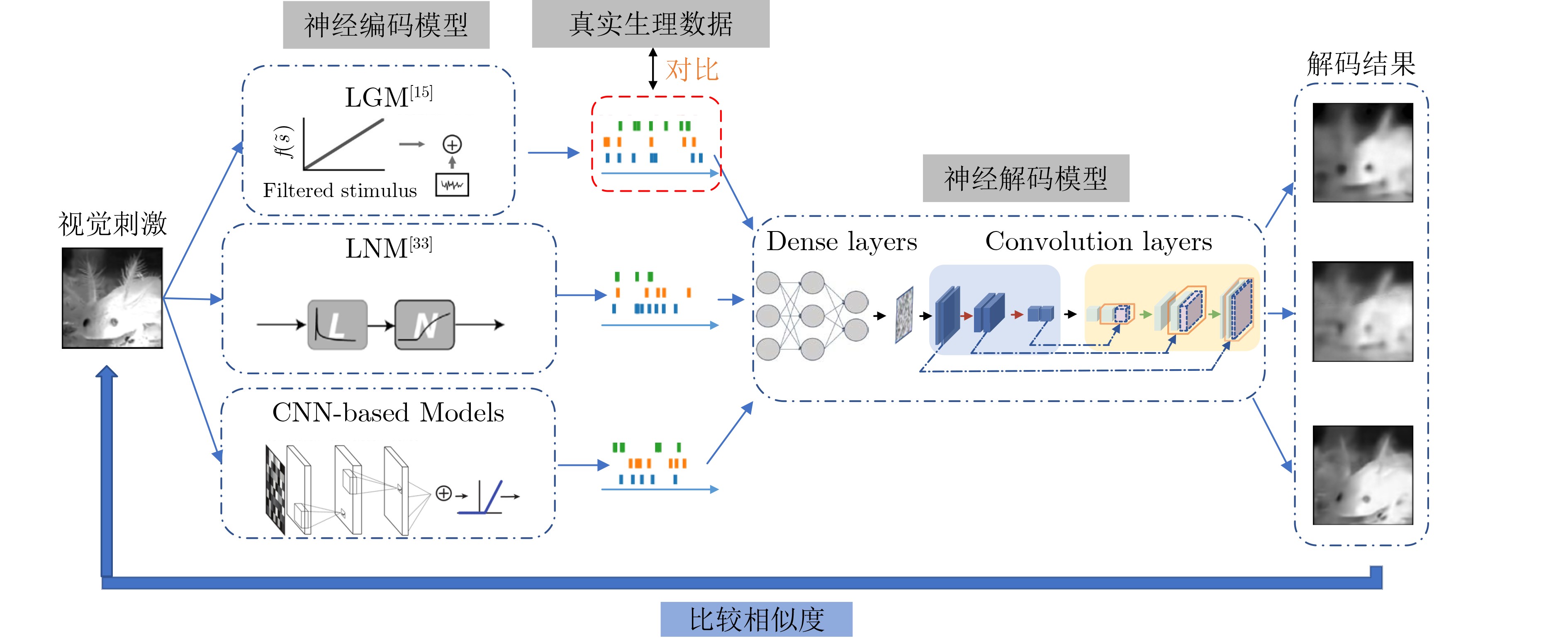
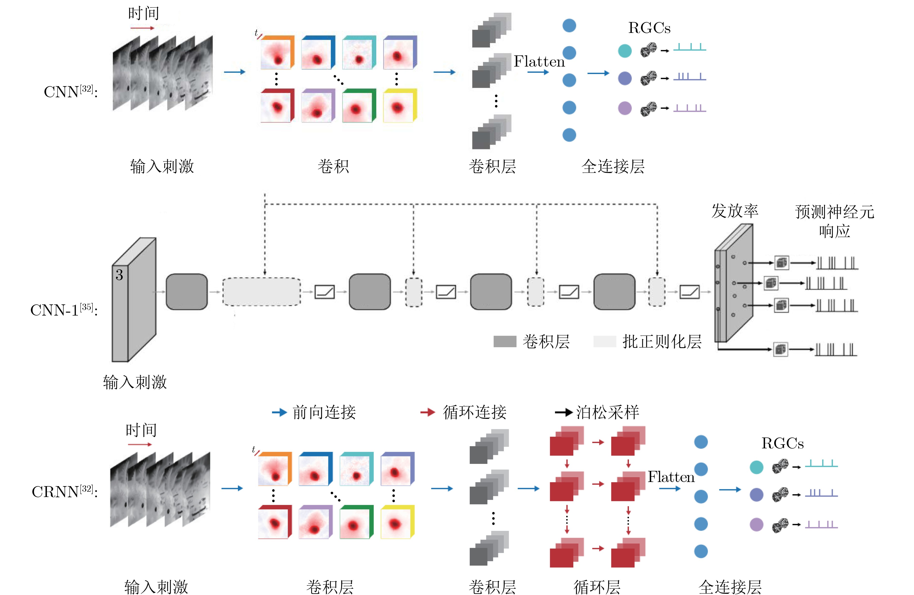
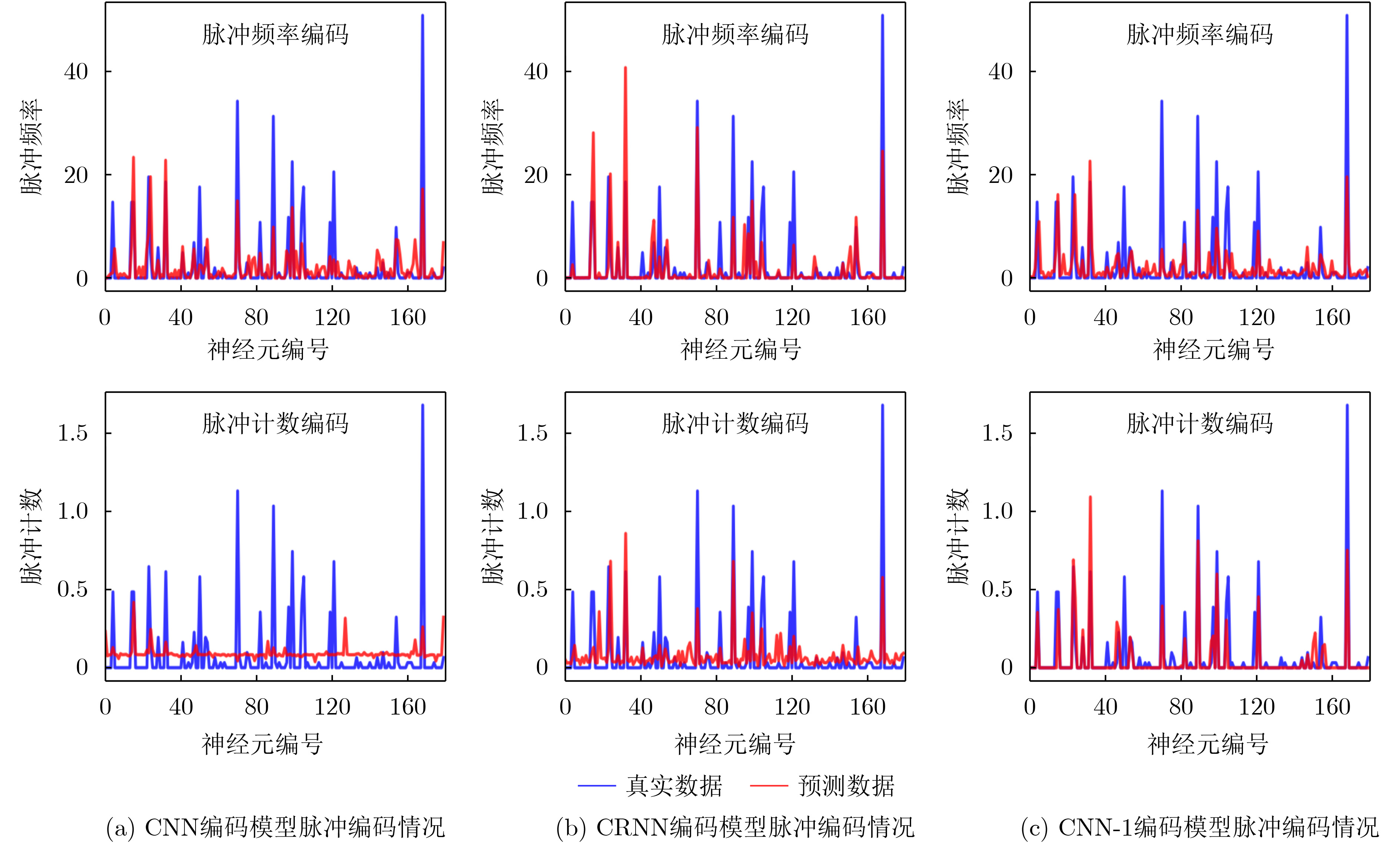
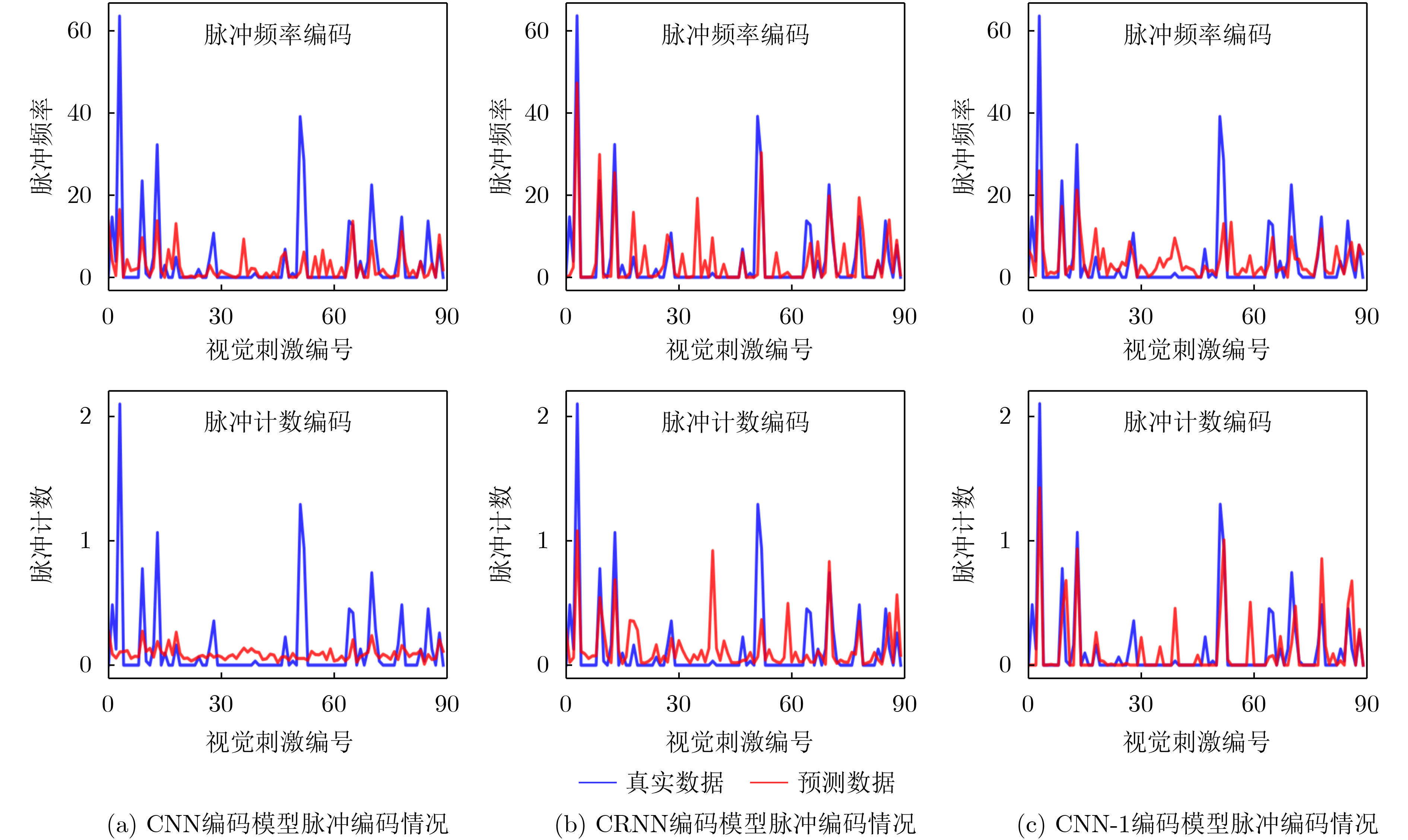
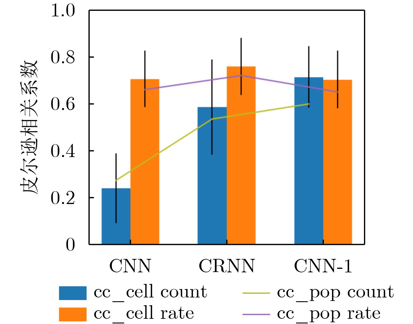
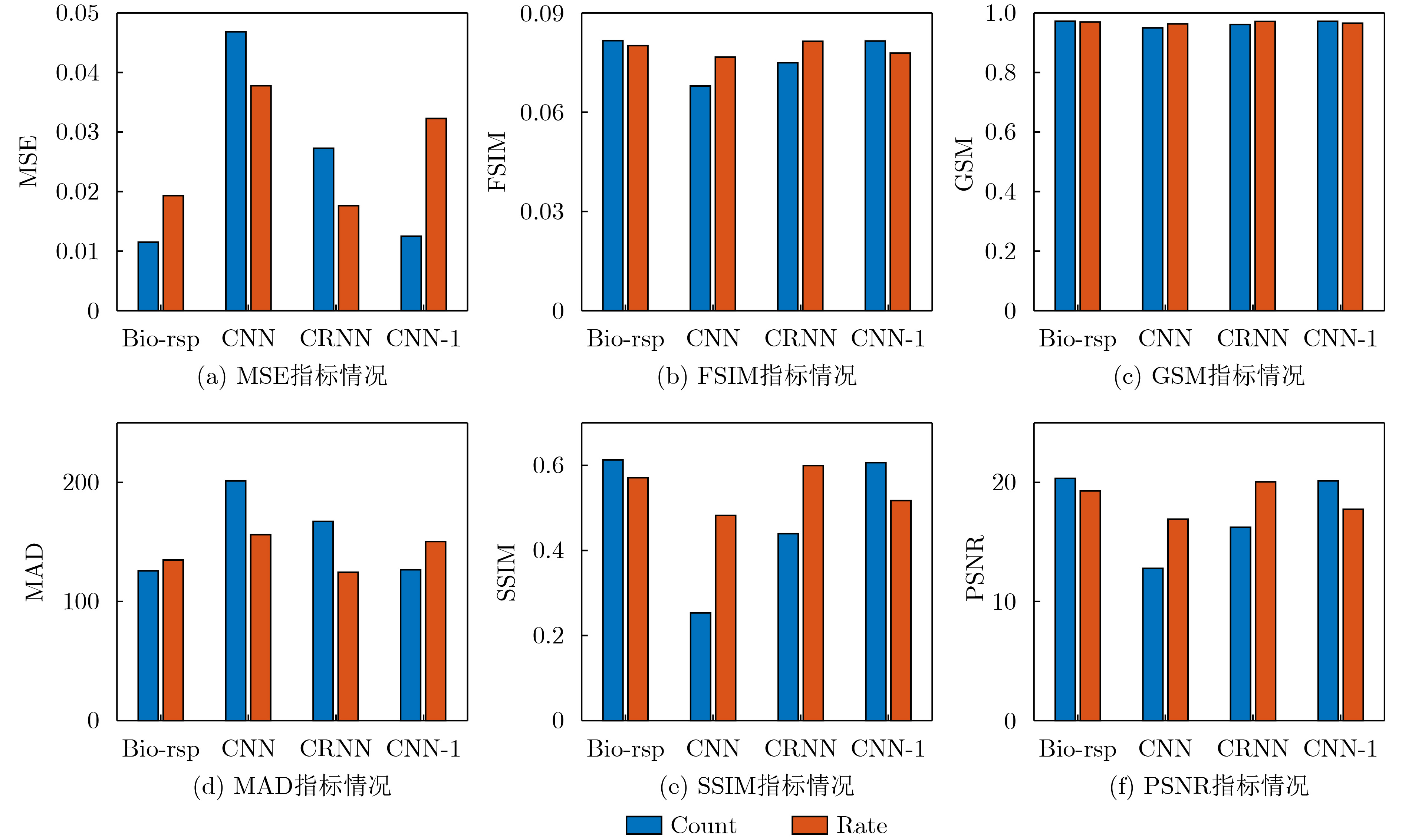
摘要: 研究界对如何对神经编码模型的性能度量还没有达成一个统一的评价标准。现有的主要编码度量方法是对神经编码模型的编码响应与真实生理响应之间的相似度进行度量。该文提出一种通过神经解码验证神经编码模型性能的方法。基于此方法构建了包括传统编码度量方法和神经解码度量方法的视觉脉冲信号编码验证框架,并基于动态视觉刺激下采集的蝾螈视网膜神经节细胞(RGC)脉冲信号数据集对此框架进行了实验验证。选择了具有动态视觉刺激脉冲响应编码能力的编码模型与性能先进的神经解码模型作为标准度量模型。实验从不同神经编码方式和不同维度全面地对3种神经编码模型的编码性能进行了度量。此外,实验结果表明,脉冲频率编码和脉冲计数编码两种编码方式对脉冲编码性能存在不可忽略的影响。Abstract: A widely recognized evaluation standard has not been reached on how to evaluate the performance of neural encoding models. Most current neural encoding evaluation methods are based on the measurement of the similarity between the encoded responses from neural encoding models and the real physiological responses. A method to validate the performance of neural encoding models through neural decoding is proposed. Using this method, a visual encoding validation framework including traditional metrics and the proposed method is constructed and experimentally validated based on a physiological dataset of Retinal Ganglion Cell (RGC) spike signals collected from salamanders over dynamic visual stimuli. Three neural encoding models with the capability of encoding the spike responses of dynamic visual stimuli and a neural decoding model with advanced performance are selected as the standard decoding models. The experiments comprehensively measure the neural encoding performance of the three neural encoding models in terms of different neural encoding methods from different perspectives. In addition, the experimental results show that there are non-negligible effects of the two neural encoding methods, i.e., rate coding and spike count coding, on the neural encoding performance.
-
Key words:
- Neural encoding /
- Neural decoding /
- Visual system /
- Brain-inspired vision
-
[1] GOODALE M A. Lessons from human vision for robotic design[J]. Autonomous Intelligent Systems, 2021, 1(1): 2. doi: 10.1007/s43684-021-00002-2 [2] GOLLISCH T and MEISTER M. Eye smarter than scientists believed: Neural computations in circuits of the retina[J]. Neuron, 2010, 65(2): 150–164. doi: 10.1016/j.neuron.2009.12.009 [3] JADZINSKY P D and BACCUS S A. Transformation of visual signals by inhibitory interneurons in retinal circuits[J]. Annual Review of Neuroscience, 2013, 36: 403–428. doi: 10.1146/annurev-neuro-062012-170315 [4] WENSEL T G. Phosphoinositides in retinal function and disease[J]. Cells, 2020, 9(4): 866. doi: 10.3390/cells9040866 [5] O’BRIEN J and BLOOMFIELD S A. Plasticity of retinal gap junctions: Roles in synaptic physiology and disease[J]. Annual Review of Vision Science, 2018, 4: 79–100. doi: 10.1146/annurev-vision-091517-034133 [6] TULI S, DASGUPTA I, GRANT E, et al. Are convolutional neural networks or transformers more like human vision?[EB/OL].https://arxiv.org/abs/2105.07197, 2021. [7] HU Yueyu, YANG Shuai, YANG Wenhan, et al. Towards coding for human and machine vision: A scalable image coding approach[C]. Proceedings of 2020 IEEE International Conference on Multimedia and Expo, London, United Kindom, 2020: 1–6. [8] LIU J K and GOLLISCH T. Spike-triggered covariance analysis reveals phenomenological diversity of contrast adaptation in the retina[J]. PLoS Computational Biology, 2015, 11(7): e1004425. doi: 10.1371/journal.pcbi.1004425 [9] LIU J K, SCHREYER H M, ONKEN A, et al. Inference of neuronal functional circuitry with spike-triggered non-negative matrix factorization[J]. Nature Communications, 2017, 8(1): 149. doi: 10.1038/s41467-017-00156-9 [10] LIU J K, KARAMANLIS D, and GOLLISCH T. Simple model for encoding natural images by retinal ganglion cells with nonlinear spatial integration[J]. bioRxiv, 2021. [11] MEYER A F, WILLIAMSON R S, LINDEN J F, et al. Models of neuronal stimulus-response functions: Elaboration, estimation, and evaluation[J]. Frontiers in Systems Neuroscience, 2017, 10: 109. doi: 10.3389/fnsys.2016.00109 [12] PILLOW J W, SHLENS J, PANINSKI L, et al. Spatio-temporal correlations and visual signalling in a complete neuronal population[J]. Nature, 2008, 454(7207): 995–999. doi: 10.1038/nature07140 [13] YAN Boyuan and NIRENBERG S. An embedded real-time processing platform for optogenetic neuroprosthetic applications[J]. IEEE Transactions on Neural Systems and Rehabilitation Engineering, 2018, 26(1): 233–243. doi: 10.1109/tnsre.2017.2763130 [14] YAN Qi, ZHENG Yajing, JIA Shanshan, et al. Revealing fine structures of the retinal receptive field by deep-learning networks[J]. IEEE Transactions on Cybernetics, 2022, 52(1): 39–50. doi: 10.1109/tcyb.2020.2972983 [15] SAHANI M and LINDEN J F. How linear are auditory cortical responses?[C]. Proceedings of the 15th International Conference on Neural Information Processing Systems, Vancouver, Canada, 2002: 125–132. [16] MCFARLAND J M, CUI Yuwei, and BUTTS D A. Inferring nonlinear neuronal computation based on physiologically plausible inputs[J]. PLoS Computational Biology, 2013, 9(7): e1003143. doi: 10.1371/journal.pcbi.1003143 [17] LINDSAY G W. Convolutional neural networks as a model of the visual system: Past, present, and future[J]. Journal of Cognitive Neuroscience, 2021, 33(10): 2017–2031. doi: 10.1162/jocn_a_01544 [18] MCINTOSH L T, MAHESWARANATHAN N, NAYEBI A, et al. Deep learning models of the retinal response to natural scenes[C]. Proceedings of the 30th International Conference on Neural Information Processing Systems, Barcelona, Spain, 2016: 1361–1369. [19] RICHARDS B A, LILLICRAP T P, BEAUDOIN P, et al. A deep learning framework for neuroscience[J]. Nature Neuroscience, 2019, 22(11): 1761–1770. doi: 10.1038/s41593-019-0520-2 [20] BATTY E, MEREL J, BRACKBILL N, et al. Multilayer recurrent network models of primate retinal ganglion cell responses[C]. Proceedings of the 5th International Conference on Learning Representations, Toulon, France, 2017. [21] MAHESWARANATHAN N, MCINTOSH L, KASTNER D B, et al. Deep learning models reveal internal structure and diverse computations in the retina under natural scenes[J]. bioRxiv, 2018: 340943. [22] YAMINS D L K and DICARLO J J. Using goal-driven deep learning models to understand sensory cortex[J]. Nature Neuroscience, 2016, 19(3): 356–365. doi: 10.1038/nn.4244 [23] 杨俊, 马正敏, 沈韬, 等. 基于深度时空特征融合的多通道运动想象EEG解码方法[J]. 电子与信息学报, 2021, 43(1): 196–203. doi: 10.11999/JEIT190300YANG Jun, MA Zhengmin, SHEN Tao, et al. Multichannel MI-EEG feature decoding based on deep learning[J]. Journal of Electronics &Information Technology, 2021, 43(1): 196–203. doi: 10.11999/JEIT190300 [24] CADENA S A, DENFIELD G H, WALKER E Y, et al. Deep convolutional models improve predictions of macaque V1 responses to natural images[J]. PLoS Computational Biology, 2019, 15(4): e1006897. doi: 10.1371/journal.pcbi.1006897 [25] YAMINS D L K, HONG Ha, CADIEU C F, et al. Performance-optimized hierarchical models predict neural responses in higher visual cortex[J]. Proceedings of the National Academy of Sciences of the United States of America, 2014, 111(23): 8619–8624. doi: 10.1073/pnas.1403112111 [26] ZHANG Yijun, YU Zhaofei, LIU J K, et al. Neural decoding of visual information across different neural recording modalities and approaches[J]. Machine Intelligence Research, 2022, 19(5): 350–365. doi: 10.1007/s11633-022-1335-2 [27] BRACKBILL N, RHOADES C, KLING A, et al. Reconstruction of natural images from responses of primate retinal ganglion cells[J]. eLife, 2020, 9: e58516. doi: 10.7554/eLife.58516 [28] WARLAND D K, REINAGEL P, and MEISTER M. Decoding visual information from a population of retinal ganglion cells[J]. Journal of Neurophysiology, 1997, 78(5): 2336–2350. doi: 10.1152/jn.1997.78.5.2336 [29] ZHANG Yichen, JIA Shanshan, ZHENG Yajing, et al. Reconstruction of natural visual scenes from neural spikes with deep neural networks[J]. Neural Networks, 2020, 125: 19–30. doi: 10.1016/j.neunet.2020.01.033 [30] ZHANG Yijun, BU Tong, ZHANG Jiyuan, et al. Decoding pixel-level image features from two-photon calcium signals of macaque visual cortex[J]. Neural Computation, 2022, 34(6): 1369–1397. doi: 10.1162/neco_a_01498 [31] XU Qi, SHEN Jiangrong, RAN Xuming, et al. Robust transcoding sensory information with neural spikes[J]. IEEE Transactions on Neural Networks and Learning Systems, 2022, 33(5): 1935–1946. doi: 10.1109/tnnls.2021.3107449 [32] ZHANG Yimeng, LEE T S, LI Ming, et al. Convolutional neural network models of V1 responses to complex patterns[J]. Journal of Computational Neuroscience, 2019, 46(1): 33–54. doi: 10.1007/s10827-018-0687-7 [33] ZHENG Yajing, JIA Shanshan, YU Zhaofei, et al. Unraveling neural coding of dynamic natural visual scenes via convolutional recurrent neural networks[J]. Patterns, 2021, 2(10): 100350. doi: 10.1016/j.patter.2021.100350 [34] SHAH N P, BRACKBILL N, RHOADES C, et al. Inference of nonlinear receptive field subunits with spike-triggered clustering[J]. eLife, 2020, 9: e45743. doi: 10.7554/elife.45743 [35] HOCHREITER S and SCHMIDHUBER J. Long short-term memory[J]. Neural Computation, 1997, 9(8): 1735–1780. doi: 10.1162/neco.1997.9.8.1735 [36] SHAH N P, BRACKBILL N, SAMARAKOON R, et al. Individual variability of neural computations in the primate retina[J]. Neuron, 2022, 110(4): 698–708.e5. doi: 10.1016/j.neuron.2021.11.026 [37] DUMOULIN V, SHLENS J, and KUDLUR M. A learned representation for artistic style[C]. Proceedings of the 5th International Conference on Learning Representations, Toulon, France, 2017. [38] WANG Zhou, BOVIK A C, SHEIKH H R, et al. Image quality assessment: From error visibility to structural similarity[J]. IEEE Transactions on Image Processing, 2004, 13(4): 600–612. doi: 10.1109/tip.2003.819861 [39] LARSON E C and CHANDLER D M. Most apparent distortion: Full-reference image quality assessment and the role of strategy[J]. Journal of Electronic Imaging, 2010, 19(1): 011006. doi: 10.1117/1.3267105 [40] ZHANG Lin, ZHANG Lei, MOU Xuanqin, et al. FSIM: A feature similarity index for image quality assessment[J]. IEEE Transactions on Image Processing, 2011, 20(8): 2378–2386. doi: 10.1109/tip.2011.2109730 [41] MORRONE M C, ROSS J, BURR D C, et al. Mach bands are phase dependent[J]. Nature, 1986, 324(6094): 250–253. doi: 10.1038/324250a0 [42] LIU Anmin, LIN Weisi, and NARWARIA M. Image quality assessment based on gradient similarity[J]. IEEE Transactions on Image Processing, 2012, 21(4): 1500–1512. doi: 10.1109/tip.2011.2175935 -

 下载:
下载:


图(8)
计量
- 文章访问数: 1114
- HTML全文浏览量: 1073
- PDF下载量: 78
- 被引次数: 0
