

EEG Source Imaging Based on fMRI Functional Network and Bayesian Matrix Decomposition
-
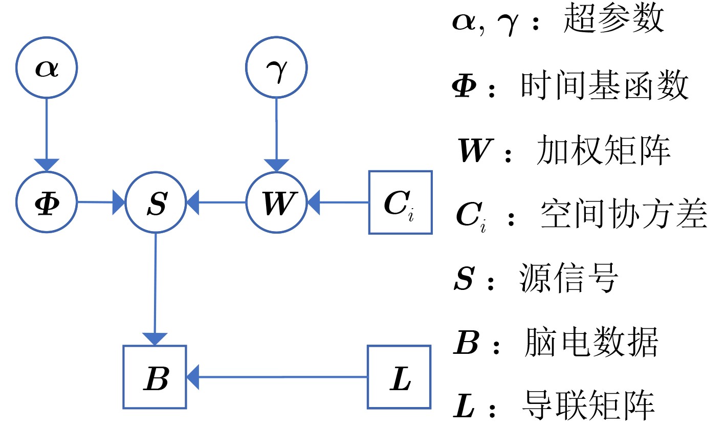
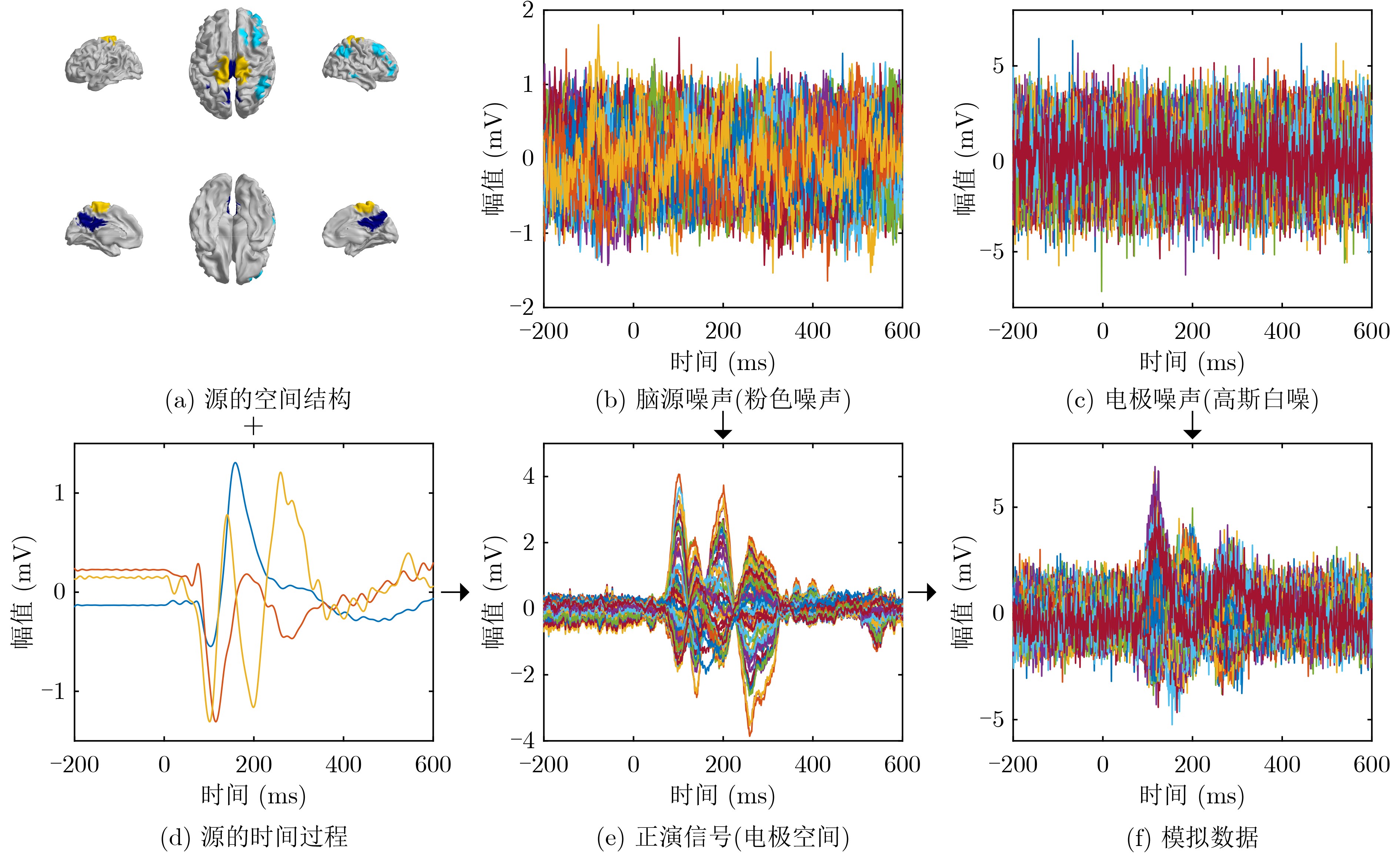
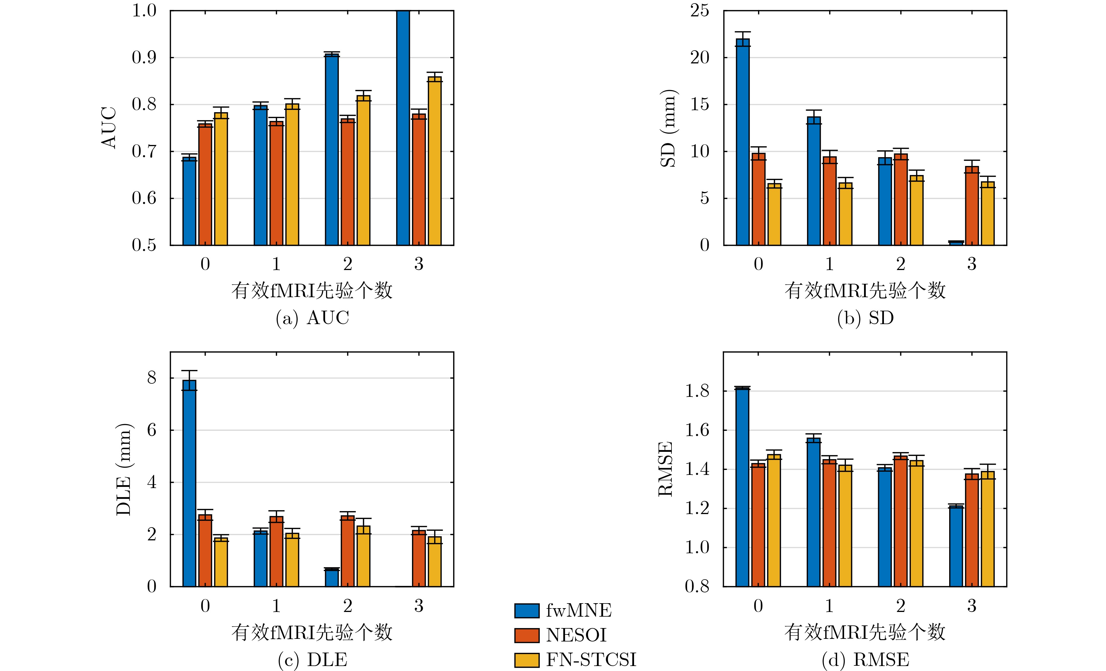
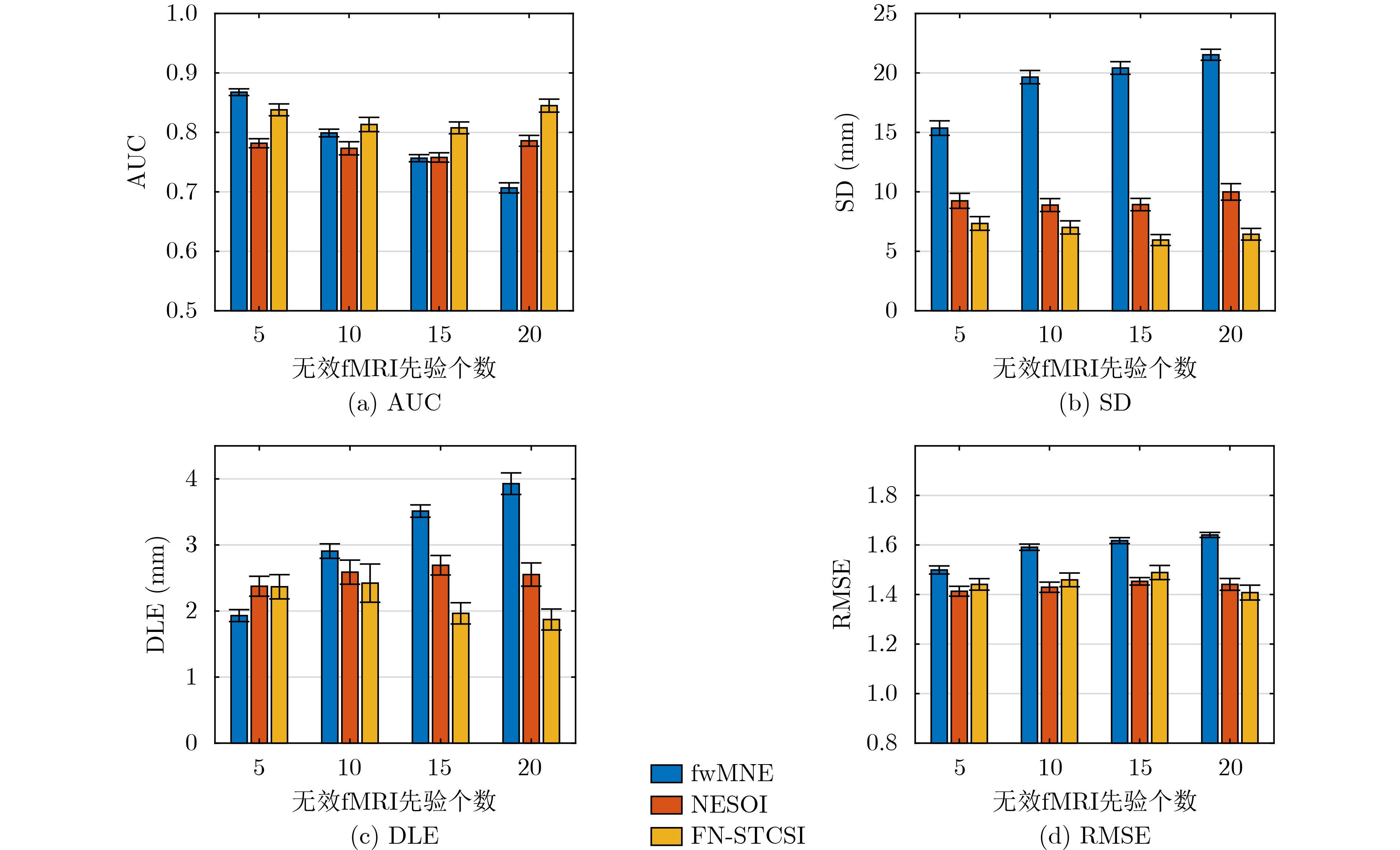
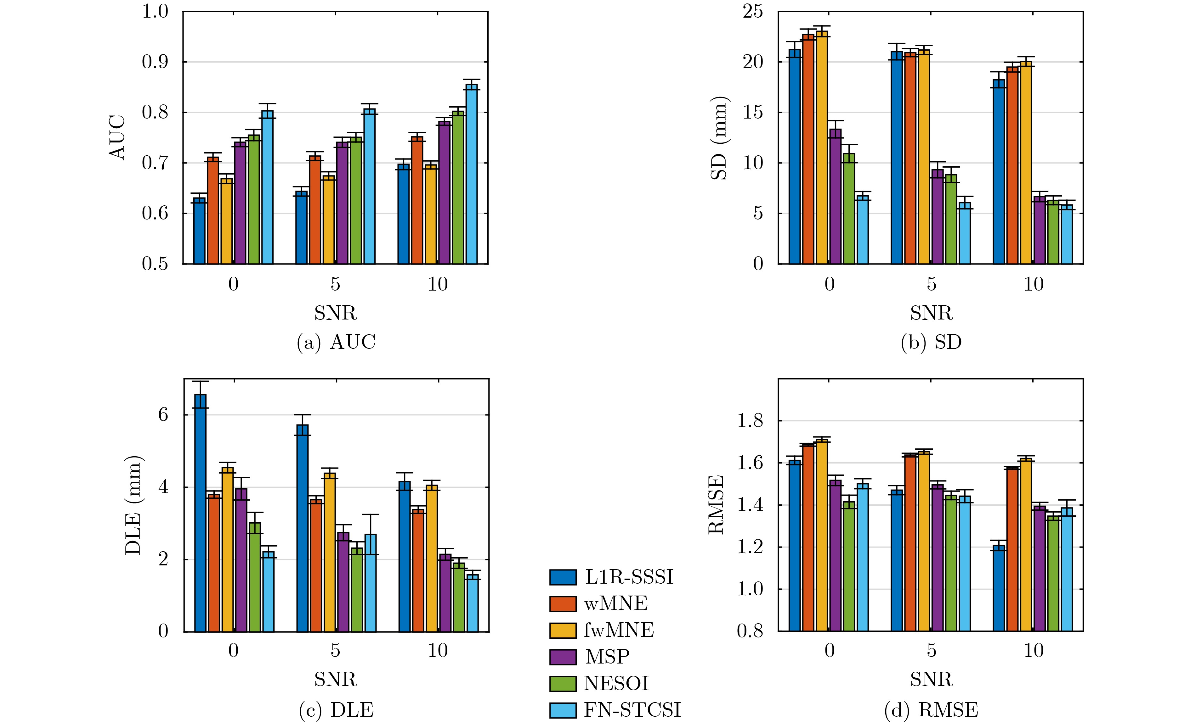
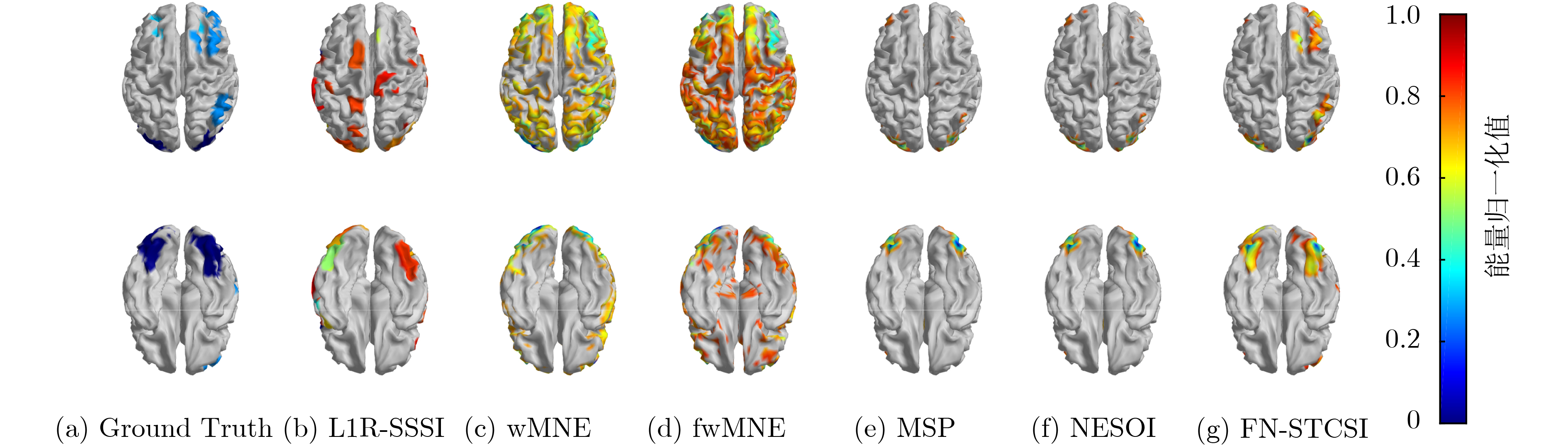
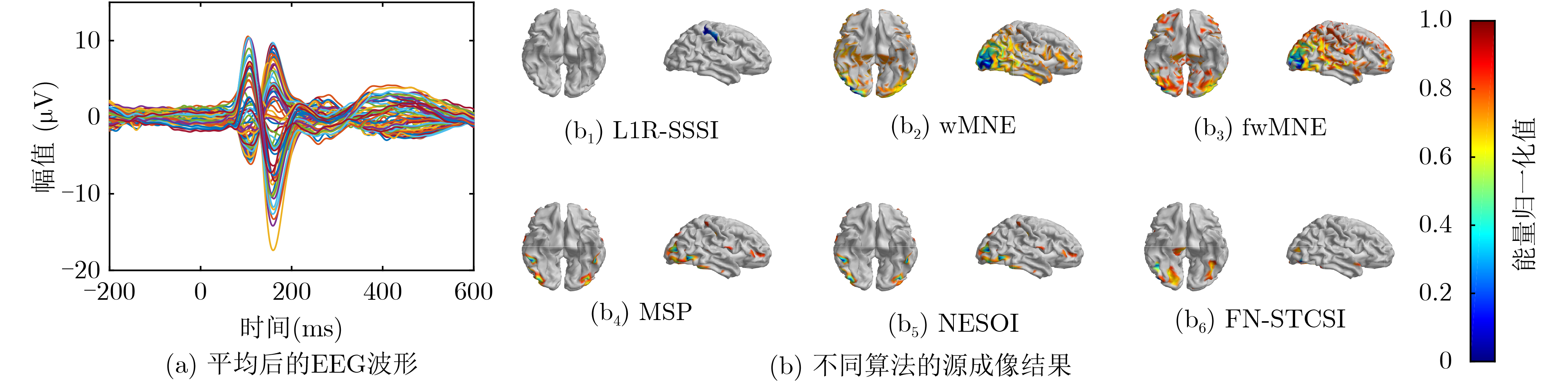
摘要: 脑电(EEG)是一种重要的脑功能成像技术,根据头皮记录的EEG信号重构皮层脑活动称为EEG源成像。然而脑源活动位置和尺寸的准确重构依然是一个挑战。为充分利用EEG和功能磁共振(fMRI)信号在时空分辨率上的互补信息,该文提出一个新的源成像方法——基于fMRI脑网络和时空约束的EEG源重构算法(FN-STCSI)。该方法在参数贝叶斯框架下,基于矩阵分解思想将源信号分解为若干时间基函数的线性组合。此外,为融合fMRI的高空间分辨率信息,FN-STCSI利用独立成分分析提取fMRI信号的功能网络,构建EEG源成像的空间协方差基,通过变分贝叶斯推断技术确定每个空间协方差基的相对贡献,实现EEG-fMRI融合。通过蒙特卡罗数值仿真和实验数据分析比较了FN-STCSI与现有算法在不同信噪比和不同先验条件下的性能,结果表明FN-STCSI能有效融合EEG-fMRI在时空上的互补信息,提高EEG弥散源成像的性能。Abstract: ElectroEncephaloGraphy (EEG) is an important brain functional imaging technology. The task to reconstruct cortical activities based on the scalp EEG is called EEG source imaging. However, the accurate reconstruction of the locations and sizes of brain source activity remains a challenge. To employ fully the spatiotemporal complementary information of EEG and functional Magnetic Resonance Imaging (fMRI), a new EEG source imaging algorithm, i.e., FN-STCSI (Functional Network based Spatio-Temporal Constrains Source Imaging) is proposed. Specifically, to make full use of the temporal information of EEG signals, the source signal matrix is decomposed into a linear combination of several time basis functions based on the idea of matrix decomposition. Additionally, to fuse the high spatial resolution information of fMRI, FN-STCSI employes independent component analysis to extract the fMRI functional networks. Then these fMRI networks are used to construct the spatial covariance basis for EEG source imaging. Variational Bayesian inference techniques are used to determine the relative contribution of each spatial covariance basis to realize EEG-fMRI fusion. Through Monte Carlo numerical simulation and experimental data analysis, FN-STCSI is compared with existing algorithms under different signal-to-noise ratios and different prior conditions. The results show that FN-STCSI can effectively fuse the complementary spatiotemporal information of EEG-fMRI and improve the performance of EEG extended source imaging.
-
[1] 张杨松, 卓彦, 尧德中. 脑电磁成像进展及展望[J]. 中国科学:生命科学, 2020, 50(11): 1268–1284. doi: 10.1360/ssv-2019-0097ZHANG Yangsong, ZHUO Yan, and YAO Dezhong. Progresses and prospects of brain electromagnetic imaging[J]. Scientia Sinica Vitae, 2020, 50(11): 1268–1284. doi: 10.1360/ssv-2019-0097 [2] OJEDA A, KREUTZ-DELGADO K, and MULLEN T. Fast and robust Block-Sparse Bayesian learning for EEG source imaging[J]. NeuroImage, 2018, 174: 449–462. doi: 10.1016/j.neuroimage.2018.03.048 [3] HE Bin, SOHRABPOUR A, BROWN E, et al. Electrophysiological source imaging: A noninvasive window to brain dynamics[J]. Annual Review of Biomedical Engineering, 2018, 20: 171–196. doi: 10.1146/annurev-bioeng-062117-120853 [4] HÄMÄLÄINEN M S and ILMONIEMI R J. Interpreting magnetic fields of the brain: Minimum norm estimates[J]. Medical & Biological Engineering & Computing, 1994, 32(1): 35–42. doi: 10.1007/BF02512476 [5] DALE A M and SERENO M I. Improved localizadon of cortical activity by combining EEG and MEG with MRI cortical surface reconstruction: A linear approach[J]. Journal of Cognitive Neuroscience, 1993, 5(2): 162–176. doi: 10.1162/jocn.1993.5.2.162 [6] GRECH R, CASSAR T, MUSCAT J, et al. Review on solving the inverse problem in EEG source analysis[J]. Journal of NeuroEngineering and Rehabilitation, 2008, 5(1): 25. doi: 10.1186/1743-0003-5-25 [7] PASCUAL-MARQUI R D. Standardized low-resolution brain electromagnetic tomography (sLORETA): Technical details[J]. Methods and Findings in Experimental and Clinical Pharmacology, 2002, 24(Suppl D): 5–12. [8] FRISTON K, HARRISON L, DAUNIZEAU J, et al. Multiple sparse priors for the M/EEG inverse problem[J]. NeuroImage, 2008, 39(3): 1104–1120. doi: 10.1016/j.neuroimage.2007.09.048 [9] CHOWDHURY R A, LINA J M, KOBAYASHI E, et al. MEG source localization of spatially extended generators of epileptic activity: Comparing entropic and hierarchical bayesian approaches[J]. PLoS One, 2013, 8(2): e55969. doi: 10.1371/journal.pone.0055969 [10] XU Furong, LIU Ke, YU Zhuliang, et al. EEG extended source imaging with structured sparsity and L1-norm residual[J]. Neural Computing and Applications, 2021, 33(14): 8513–8524. doi: 10.1007/s00521-020-05603-1 [11] 周伊婕, 宋西姊, 何峰, 等. 基于脑电的多模态神经功能成像新技术研究进展[J]. 中国生物医学工程学报, 2020, 39(5): 595–602. doi: 10.3969/j.issn.0258-8021.2020.05.010ZHOU Yijie, SONG Xizi, HE Feng, et al. Research progress of multimodal functional neural imaging technology based on EEG[J]. Chinese Journal of Biomedical Engineering, 2020, 39(5): 595–602. doi: 10.3969/j.issn.0258-8021.2020.05.010 [12] LIU A K, BELLIVEAU J W, and DALE A M. Spatiotemporal imaging of human brain activity using functional MRI constrained magnetoencephalography data: Monte Carlo simulations[J]. Proceedings of the National Academy of Sciences of the United States of America, 1998, 95(15): 8945–8950. doi: 10.1073/pnas.95.15.8945 [13] HENSON R N, FLANDIN G, FRISTON K J, et al. A parametric empirical Bayesian framework for fMRI-constrained MEG/EEG source reconstruction[J]. Human Brain Mapping, 2010, 31(10): 1512–1531. doi: 10.1002/hbm.20956 [14] LEI Xu, XU Peng, LUO Cheng, et al. fMRI functional networks for EEG source imaging[J]. Human Brain Mapping, 2011, 32(7): 1141–1160. doi: 10.1002/hbm.21098 [15] ZUMER J M, ATTIAS H T, SEKIHARA K, et al. Probabilistic algorithms for MEG/EEG source reconstruction using temporal basis functions learned from data[J]. NeuroImage, 2008, 41(3): 924–940. doi: 10.1016/j.neuroimage.2008.02.006 [16] OU Wanmei, HÄMÄLÄINEN M S, and GOLLAND P. A distributed spatio-temporal EEG/MEG inverse solver[J]. Neuroimage, 2009, 44(3): 932–946. doi: 10.1016/j.neuroimage.2008.05.063 [17] LIU Ke, YU Zhuliang, WU Wei, et al. Bayesian electromagnetic spatio-temporal imaging of extended sources based on matrix factorization[J]. IEEE Transactions on Biomedical Engineering, 2019, 66(9): 2457–2469. doi: 10.1109/TBME.2018.2890291 [18] ENGEMANN D A and GRAMFORT A. Automated model selection in covariance estimation and spatial whitening of MEG and EEG signals[J]. NeuroImage, 2015, 108: 328–342. doi: 10.1016/j.neuroimage.2014.12.040 [19] TRUJILLO-BARRETO N J, AUBERT-VÁZQUEZ E, and PENNY W D. Bayesian M/EEG source reconstruction with spatio-temporal priors[J]. NeuroImage, 2008, 39(1): 318–335. doi: 10.1016/j.neuroimage.2007.07.062 [20] HENSON R N, ABDULRAHMAN H, FLANDIN G, et al. Multimodal integration of M/EEG and f/MRI data in SPM12[J]. Frontiers in Neuroscience, 2019, 13: 300. doi: 10.3389/fnins.2019.00300 [21] OTSU N. A threshold selection method from gray-level histograms[J]. IEEE Transactions on Systems, Man, and Cybernetics, 1979, 9(1): 62–66. doi: 10.1109/TSMC.1979.4310076 [22] ASADZADEH S, YOUSEFI REZAII T, BEHESHTI S, et al. A systematic review of EEG source localization techniques and their applications on diagnosis of brain abnormalities[J]. Journal of Neuroscience Methods, 2020, 339: 108740. doi: 10.1016/j.jneumeth.2020.108740 [23] AL-SAFFAR A, BIALKOWSKI A, BAKTASHMOTLAGH M, et al. Closing the gap of simulation to reality in electromagnetic imaging of brain strokes via deep neural networks[J]. IEEE Transactions on Computational Imaging, 2021, 7: 13–21. doi: 10.1109/tci.2020.3041092 [24] BORE J C, LI P, JIANG L, et al. A long short-term memory network for sparse spatiotemporal EEG source imaging[J]. IEEE Transactions on Medical Imaging, 2021, 40(12): 3787–3800. doi: 10.1109/TMI.2021.3097758 [25] HOU Yimin, ZHOU Lu, JIA Shuyue, et al. A novel approach of decoding EEG four-class motor imagery tasks via scout ESI and CNN[J]. Journal of Neural Engineering, 2020, 17(1): 016048. doi: 10.1088/1741-2552/ab4af6 [26] WU Wei, ZHANG Yu, JIANG Jing, et al. An electroencephalographic signature predicts antidepressant response in major depression[J]. Nature Biotechnology, 2020, 38(4): 439–447. doi: 10.1038/s41587-019-0397-3 [27] TOLL R T, WU Wei, NAPARSTEK S, et al. An electroencephalography connectomic profile of posttraumatic stress disorder[J]. American Journal of Psychiatry, 2020, 177(3): 233–243. doi: 10.1176/appi.ajp.2019.18080911 -

 下载:
下载:


图(8)
计量
- 文章访问数: 1389
- HTML全文浏览量: 815
- PDF下载量: 152
- 被引次数: 0
