

Virtual Reality Motion Sickness Recognition Model Driven by Lead-attention and Brain Connection
-
摘要: 虚拟现实晕动症(VRMS)是阻碍虚拟现实技术行业发展的重要问题,检测VRMS水平是研究并克服这一问题的先决条件。所以该文引入并改进了一种脑电端到端识别模型定量识别用户在使用虚拟现实时的VRMS水平。该模型首先利用1维卷积神经网络(CNN)对脑电信号进行滤波,然后计算导联间相关性构成功能脑网络,最后利用CNN和全连接层提取脑网络特征和回归分析。该文通过优化1维卷积核大小及加入一种新型导联注意力结构来增强该模型特征提取能力。最后采用虚拟现实场景《VRQ test》诱发受试者产生VRMS并记录受试者脑电信号及主观评价VRMS水平(模拟器眩晕量表SSQ),所得数据用于验证该模型。结果显示经过10折交叉验证该方法检测到的VRMS水平与真实值之间平均均方误差为15.10,平均拟合优度为:96.63%。该结果表明该文所提模型可用于虚拟现实晕动症的检测,该脑电检测方法有望成为一种通用的虚拟现实产品评估方法。
-
关键词:
- 虚拟现实晕动症(VRMS) /
- 脑电(EEG) /
- 功能脑网络 /
- 导联注意力 /
- 模拟器眩晕量表(SSQ)
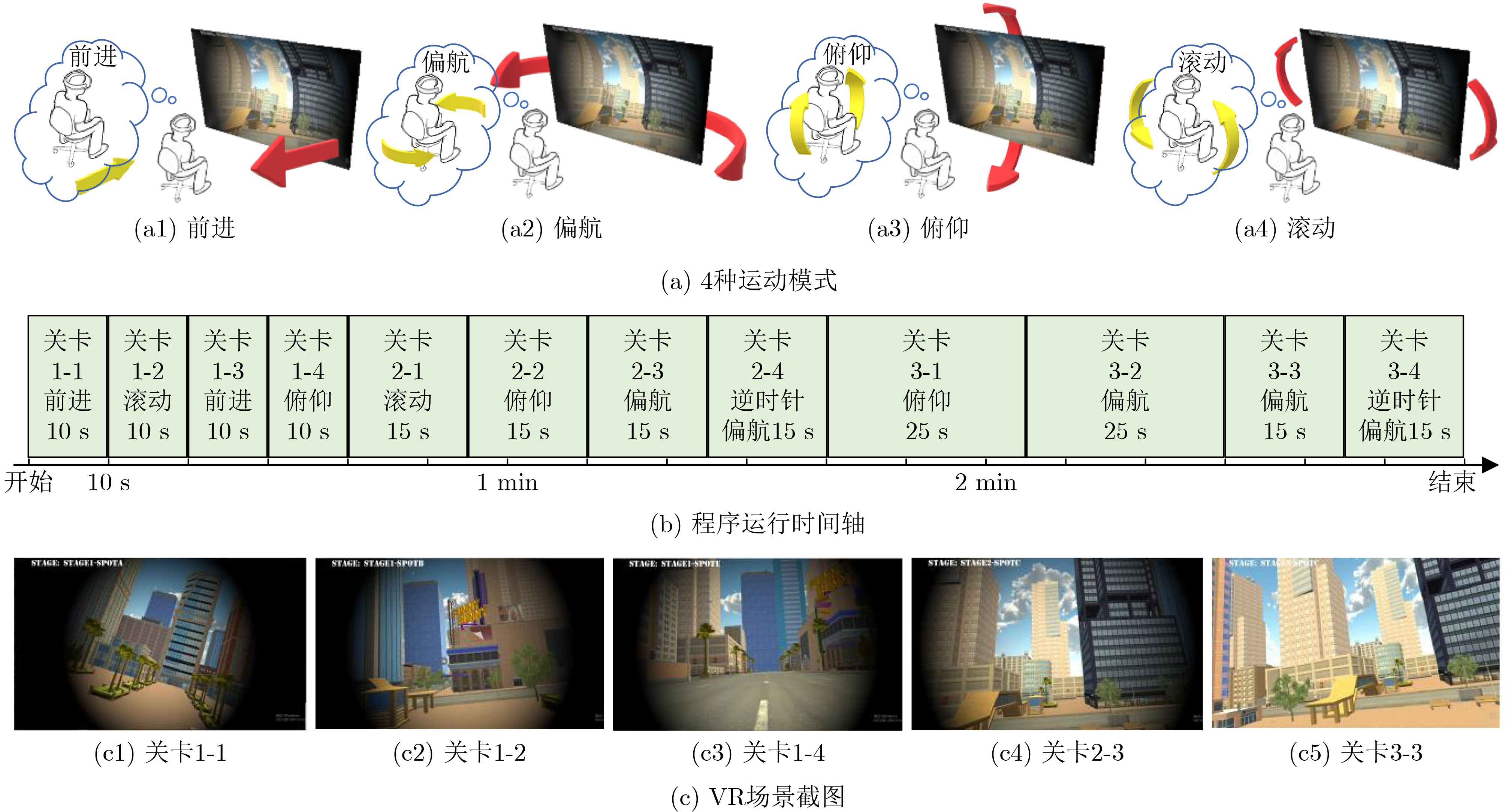
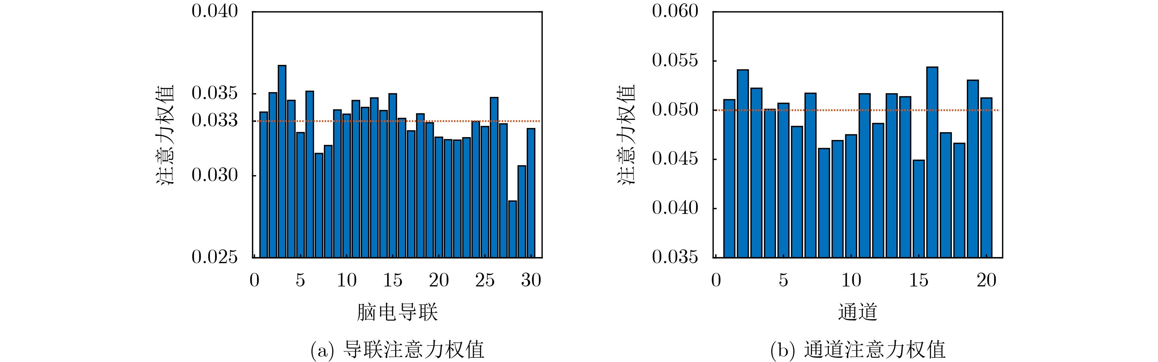
Abstract:Objective Virtual Reality Motion Sickness (VRMS) hinders the development of virtual reality technology and affects users’ experience, potentially threatening their health. Accurately assessing VRMS levels is essential for studying its causes and treatment strategies. ElectroEncephaloGram (EEG) provides a non-invasive, low-cost method with high temporal resolution, reflecting real-time neural activity, making it suitable for VRMS assessment. This paper introduces and improves an end-to-end EEG regression model based on Convolutional Neural Networks (CNN) and functional brain networks, termed Brain Connection-based CNN (BCCNN), to quantitatively recognize VRMS in users within a VR environment. Methods The BCCNN utilizes a 1D-CNN to filter EEG signals and compute correlation coefficients among electrodes, forming functional brain networks. It then employs CNN and fully connected layers to extract network features and perform regression analysis ( Figure 2 ). This study optimizes the kernel size of the 1D-CNN and proposes a novel lead attention module to enhance feature extraction. The attention module, inspired by the squeeze-and-excitation mechanism, computes the weights from the filtered EEG signals rather than the extracted features. Additionally, the attention weights are derived from and applied to the leads of the EEG (Figure 3 ). To induce VRMS, a virtual reality scene called “VRQ test” is used. The subject’s EEG signal and subjective VRMS level, recorded via the Simulator Sickness Questionnaire (SSQ), are collected. These data are then used to validate the model (Figure 2 ). The model’s performance is evaluated using Mean Squared Error (MSE), Mean Absolute Error (MAE), and the goodness of fit (R2), comparing the predicted VRMS levels with the real values. A comparison with reference methods is conducted to assess the effectiveness of the BCCNN and its optimizations.Results and Discussions The results show that the optimized kernel size of the 1D-CNN layer is 16, reducing the average MSE of the original BCCNN by 6.53 ( Table 1 ,Table 2 ,Figure 4 ). Additionally, the lead attention module further improves the BCCNN, lowering the average MSE by 7.65 compared to the original model, outperforming the channel attention module (Table 2 ). The optimized BCCNN achieves an average MSE of 15.10 and an average R2 of 96.63% in 10-fold cross-validation, significantly exceeding the original BCCNN and ten state-of-the-art and baseline models (Table 2 ). Among the reference methods, the combination of difference entropy and Gaussian process regression yields the best performance. Furthermore, reference models using a filter bank outperform other reference models, indicating that handcrafted processing of the EEG data can enhance model performance (Table 2 ). Visualizing the functional connections and extracted features of the BCCNN reveals that functional connections are stronger at higher VRMS levels compared to lower VRMS levels.Conclusions This study introduces and optimizes the BCCNN for assessing VRMS using EEG. The main innovation of this work lies in optimizing the kernel size of the 1D-CNN and proposing a novel lead attention module. The results demonstrate that these optimizations enhance the accuracy of VRMS assessment, with the updated model offering a more precise evaluation. EEG is thus expected to become a standard method for assessing VRMS in VR products. The proposed approach enables VRMS assessment during and after a user’s experience in a VR scene. -
表 1 不同类型1D卷积核及不同类型输出层激活函数下BCCNN模型的回归表现
激活函数 卷积核长度 均方误差(MSE) 平均绝对误差(MAE) 拟合优度(R2,%) Sigmoid 最优16 18.40±3.76 2.34±0.26 95.84±0.90 最差29 25.1±5.30 2.73±0.44 94.16±1.45 线性 最优16 29.98±6.00 3.35±0.46 92.96±1.65 最差23 41.21±21.54 3.82±0.93 90.02±5.61  下载: 导出CSV
表 2 改进BCCNN模型与其他常用模型对VRMS数据的回归表现对比(Mean±std.)
# 方法 均方误差(MSE) 平均绝对误差(MAE) 拟合优度R2 (%) 1 节律能量及能量比[12]+mRMR[29]+高斯过程回归 60.50±3.68 5.13±0.13 83.86±1.74 2 微分熵[27]+mRMR+高斯过程回归 38.65±2.37 4.18±0.11 90.84±0.86 3 EMDPLV+图论指标+mRMR+高斯过程回归 154.37±14.17 9.29±0.27 50.04±5.74 4 Shallow ConvNet[30] 217.76±27.20 11.17±0.80 6.22±11.10 5 Deep ConvNet[30] 213.55±49.97 11.62±1.53 2.58±5.98 6 FBtCNN[31] 171.03±16.94 9.91±0.55 23.47±11.78 7 SSVEPNet[32] 166.29±22.23 8.91±0.69 67.24±4.05 8 1D-CNN(时间、导联)+LSTM 123.92±17.95 7.16±0.75 68.74±5.68 9 ADFCNN[33] 122.74±22.05 7.42±0.79 67.45±7.38 10 MOCNN[34] 91.74±22.28 7.17±1.02 77.38±6.63 11 Conformer[35] 82.15±9.86 5.44±0.27 80.92±2.58 12 FBCNet[36] 77.29±8.19 6.87±0.38 80.08±2.63 13 滤波器组+1D-CNN(导联)+LSTM 53.62±6.55 5.61±0.36 86.83±3.25 14 原版BCCNN 24.93±4.60 2.8±0.399 94.16±1.23 15 BCCNN+优化卷积核 18.40±3.76 2.34±0.26 95.84±0.90 16 BCCNN+通道注意力 22.73±6.49 2.25±0.32 94.90±1.39 17 BCCNN+优化卷积核+通道注意力 20.80±6.72 2.22±0.34 95.29±1.65 18 BCCNN+导联注意力 17.28±3.68 2.08±0.28 96.10±0.90 19 BCCNN+优化卷积核+导联注意力 15.10±4.32 1.93±0.28 96.63±1.09
下载: 导出CSV
-
[1] KENNEDY R S, DREXLER J, and KENNEDY R C. Research in visually induced motion sickness[J]. Applied Ergonomics, 2010, 41(4): 494–503. doi: 10.1016/j.apergo.2009.11.006. [2] NALIVAIKO E, DAVIS S L, BLACKMORE K L, et al. Cybersickness provoked by head-mounted display affects cutaneous vascular tone, heart rate and reaction time[J]. Physiology & Behavior, 2015, 151: 583–590. doi: 10.1016/j.physbeh.2015.08.043. [3] MASON B, 王麒. 虚拟现实会增加晕动病的风险[J]. 中国科技教育, 2017(2): 65–66.MASON B, WANG Qi. Virtual reality raises real risk of motion sickness[J]. China Science & Technology Educationb, 2017(2): 65–66. [4] 易琳, 贾瑞双, 刘然, 等. 虚拟现实环境中视觉诱导晕动症的评估指标[J]. 航天医学与医学工程, 2018, 31(4): 437–445. doi: 10.16289/j.cnki.1002–0837.2018.04.008.YI Lin, JIA Ruishuang, LIU Ran et al. Evaluation indicators for visually induced motion sickness in virtual reality environment[J]. Space Medicine & Medical Engineering, 2018, 31(4): 437–445. doi: 10.16289/j.cnki.1002–0837.2018.04.008. [5] KIM J, OH H, KIM W, et al. A deep motion sickness predictor induced by visual stimuli in virtual reality[J]. IEEE Transactions on Neural Networks and Learning Systems, 2022, 33(2): 554–566. doi: 10.1109/tnnls.2020.3028080. [6] KIM H G, LIM H T, LEE S, et al. VRSA net: VR sickness assessment considering exceptional motion for 360° VR video[J]. IEEE Transactions on Image Processing, 2019, 28(4): 1646–1660. doi: 10.1109/tip.2018.2880509. [7] WANG Yuyang, CHARDONNET J R, and MERIENNE F. VR sickness prediction for navigation in immersive virtual environments using a deep long short term memory model[C]. 2019 IEEE Conference on Virtual Reality and 3D User Interfaces (VR), Osaka, Japan, 2019: 1874–1881. doi: 10.1109/VR.2019.8798213. [8] 耿跃华, 石金祥. 机器学习与脑电信号分析相结合的眩晕状态分类[J]. 中国组织工程研究, 2022, 26(29): 4624–4631. doi: 10.12307/2022.844.GENG Yuehua and SHI Jinxiang. Classification of vertigo state based on machine learning and electroencephalogram signal analysis[J]. Chinese Journal of Tissue Engineering Research, 2022, 26(29): 4624–4631. doi: 10.12307/2022.844. [9] JANG K M, WOO Y S, and LIM H K. Electrophysiological changes in the virtual reality sickness: EEG in the VR sickness[C]. The 25th International Conference on 3D Web Technology, 2020: 26. doi: 10.1145/3424616.3424722. [10] LIM H K, JI K, WOO Y S, et al. Test-retest reliability of the virtual reality sickness evaluation using electroencephalography (EEG)[J]. Neuroscience Letters, 2021, 743: 135589. doi: 10.1016/j.neulet.2020.135589. [11] NAQVI S A A, BADRUDDIN N, JATOI M A, et al. EEG based time and frequency dynamics analysis of visually induced motion sickness (VIMS)[J]. Australasian Physical & Engineering Sciences in Medicine, 2015, 38(4): 721–729. doi: 10.1007/s13246-015-0379-9. [12] LI Xiaolu, ZHU Changrong, XU Cangsu, et al. VR motion sickness recognition by using EEG rhythm energy ratio based on wavelet packet transform[J]. Computer Methods and Programs in Biomedicine, 2020, 188: 105266. doi: 10.1016/j.cmpb.2019.105266. [13] CHEN Y C, DUANN J R, CHUANG S W, et al. Spatial and temporal EEG dynamics of motion sickness[J]. NeuroImage, 2010, 49(3): 2862–2870. doi: 10.1016/j.neuroimage.2009.10.005. [14] KROKOS E and VARSHNEY A. Quantifying VR cybersickness using EEG[J]. Virtual Reality, 2022, 26(1): 77–89. doi: 10.1007/s10055-021-00517-2. [15] WU Jintao, ZHOU Qianxiang, LI Jiaxuan, et al. Inhibition-related N2 and P3: Indicators of visually induced motion sickness (VIMS)[J]. International Journal of Industrial Ergonomics, 2020, 78: 102981. doi: 10.1016/j.ergon.2020.102981. [16] PARK S, KIM L, KWON J, et al. Evaluation of visual-induced motion sickness from head-mounted display using heartbeat evoked potential: A cognitive load-focused approach[J]. Virtual Reality, 2022, 26(3): 979–1000. doi: 10.1007/s10055-021-00600-8. [17] LIU Ran, XU Miao, ZHANG Yanzhen, et al. A pilot study on electroencephalogram-based evaluation of visually induced motion sickness[J]. Journal of Imaging Science and Technology, 2020, 64(2): 020501. doi: 10.2352/J.ImagingSci.Technol.2020.64.2.020501. [18] CECOTTI H and GRASER A. Convolutional neural networks for P300 detection with application to brain-computer interfaces[J]. IEEE Transactions on Pattern Analysis and Machine Intelligence, 2011, 33(3): 433–445. doi: 10.1109/tpami.2010.125. [19] TABAR Y R and HALICI U. A novel deep learning approach for classification of EEG motor imagery signals[J]. Journal of Neural Engineering, 2017, 14(1): 016003. doi: 10.1088/1741-2560/14/1/016003. [20] ACHARYA U R, OH S L, HAGIWARA Y, et al. Automated EEG-based screening of depression using deep convolutional neural network[J]. Computer Methods and Programs in Biomedicine, 2018, 161: 103–113. doi: 10.1016/j.cmpb.2018.04.012. [21] HU Jie, SHEN Li, ALBANIE S, et al. Squeeze-and-excitation networks[J]. IEEE Transactions on Pattern Analysis and Machine Intelligence, 2020, 42(8): 2011–2023. doi: 10.1109/tpami.2019.2913372. [22] HUANG Jing, REN Lifeng, ZHOU Xiaokang, et al. An improved neural network based on SENet for sleep stage classification[J]. IEEE Journal of Biomedical and Health Informatics, 2022, 26(10): 4948–4956. doi: 10.1109/jbhi.2022.3157262. [23] HE Yanbin, LU Zhiyang, WANG Jun, et al. A self-supervised learning based channel attention MLP-mixer network for motor imagery decoding[J]. IEEE Transactions on Neural Systems and Rehabilitation Engineering, 2022, 30: 2406–2417. doi: 10.1109/tnsre.2022.3199363. [24] NIU Weixin, MA Chao, SUN Xinlin, et al. A brain network analysis-based double way deep neural network for emotion recognition[J]. IEEE Transactions on Neural Systems and Rehabilitation Engineering, 2023, 31: 917–925. doi: 10.1109/tnsre.2023.3236434. [25] HUA Chengcheng, WANG Hong, CHEN Jichi, et al. Novel functional brain network methods based on CNN with an application in proficiency evaluation[J]. Neurocomputing, 2019, 359: 153–162. doi: 10.1016/j.neucom.2019.05.088. [26] KENNEDY R S, LANE N E, BERBAUM K S, et al. Simulator sickness questionnaire: An enhanced method for quantifying simulator sickness[J]. The International Journal of Aviation Psychology, 1993, 3(3): 203–220. doi: 10.1207/s15327108ijap0303_3. [27] ZHENG Weilong, LIU Wei, LU Yifei, et al. EmotionMeter: A multimodal framework for recognizing human emotions[J]. IEEE Transactions on Cybernetics, 2019, 49(3): 1110–1122. doi: 10.1109/tcyb.2018.2797176. [28] 苗敏敏, 徐宝国, 胡文军, 等. 基于自适应优化空频微分熵的情感脑电识别[J]. 仪器仪表学报, 2021, 42(3): 221–230. doi: 10.19650/j.cnki.cjsi.J2006936.MIAO Minmin, XU Baoguo, HU Wenjun, et al. Emotion EEG recognition based on the adaptive optimized spatial-frequency differential entropy[J]. Chinese Journal of Scientific Instrument, 2021, 42(3): 221–230. doi: 10.19650/j.cnki.cjsi.J2006936. [29] PENG Hanchuan, LONG Fuhui, and DING C. Feature selection based on mutual information criteria of max-dependency, max-relevance, and min-redundancy[J]. IEEE Transactions on Pattern Analysis and Machine Intelligence, 2005, 27(8): 1226–1238. doi: 10.1109/Tpami.2005.159. [30] SCHIRRMEISTER R T, SPRINGENBERG J T, FIEDERER L D J, et al. Deep learning with convolutional neural networks for EEG decoding and visualization[J]. Human Brain Mapping, 2017, 38(11): 5391–5420. doi: 10.1002/hbm.23730. [31] DING Wenlong, SHAN Jianhua, FANG Bin, et al. Filter bank convolutional neural network for short time-window steady-state visual evoked potential classification[J]. IEEE Transactions on Neural Systems and Rehabilitation Engineering, 2021, 29: 2615–2624. doi: 10.1109/tnsre.2021.3132162. [32] PAN Yudong, CHEN Jianbo, ZHANG Yangsong, et al. An efficient CNN-LSTM network with spectral normalization and label smoothing technologies for SSVEP frequency recognition[J]. Journal of Neural Engineering, 2022, 19(5): 056014. doi: 10.1088/1741-2552/ac8dc5. [33] TAO Wei, WANG Ze, WONG C M, et al. ADFCNN: Attention-based dual-scale fusion convolutional neural network for motor imagery brain-computer interface[J]. IEEE Transactions on Neural Systems and Rehabilitation Engineering, 2024, 32: 154–165. doi: 10.1109/TNSRE.2023.3342331. [34] JIN Jing, XU Ruitian, DALY I, et al. MOCNN: A multiscale deep convolutional neural network for ERP-based brain-computer interfaces[J]. IEEE Transactions on Cybernetics, 2024, 54(9): 5565–5576. doi: 10.1109/TCYB.2024.3390805. [35] SONG Yonghao, ZHENG Qingqing, LIU Bingchuan, et al. EEG conformer: Convolutional transformer for EEG decoding and visualization[J]. IEEE Transactions on Neural Systems and Rehabilitation Engineering, 2023, 31: 710–719. doi: 10.1109/TNSRE.2022.3230250. [36] MANE R, ROBINSON N, VINOD A P, et al. A multi-view CNN with novel variance layer for motor imagery brain computer interface[C]. 2020 42nd Annual International Conference of the IEEE Engineering in Medicine & Biology Society, Montreal, Canada, 2020: 2950–2953. doi: 10.1109/EMBC44109.2020.9175874. -

 下载:
下载:


图(6) / 表(2)
计量
- 文章访问数: 990
- HTML全文浏览量: 725
- PDF下载量: 65
- 被引次数: 0
