

An Adaptive Medical Ultrasound Images Despeckling Method Based on Deep Learning
-
摘要:
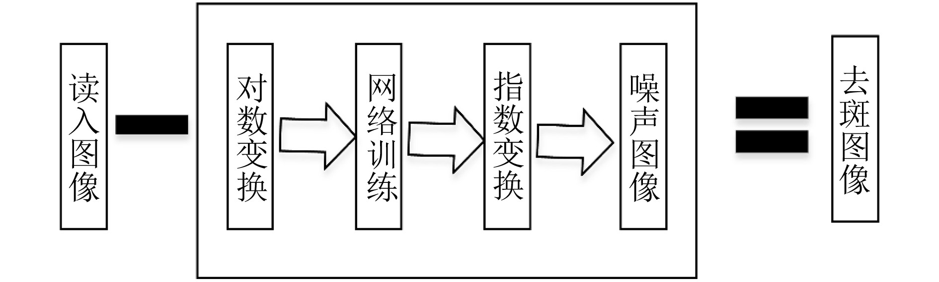
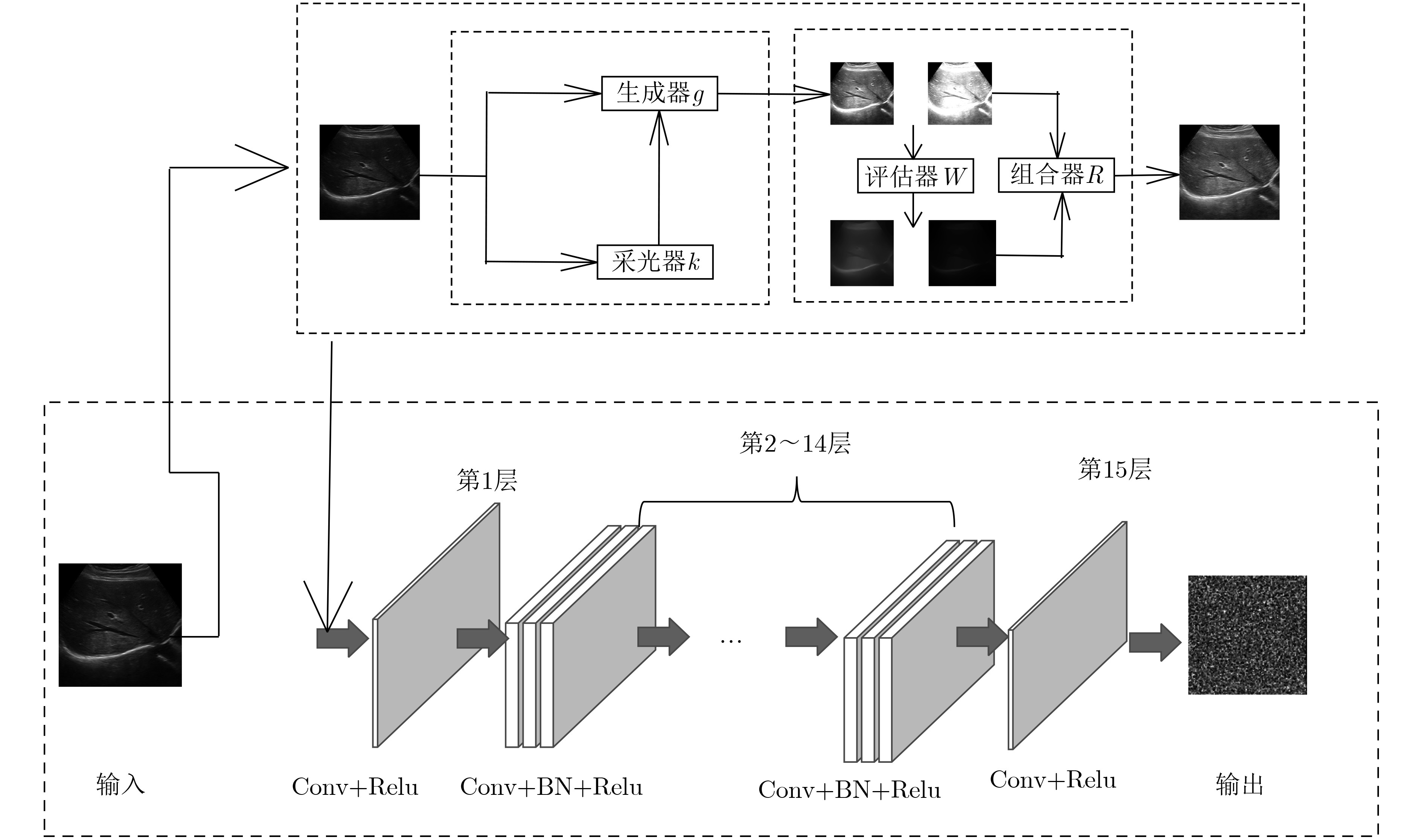
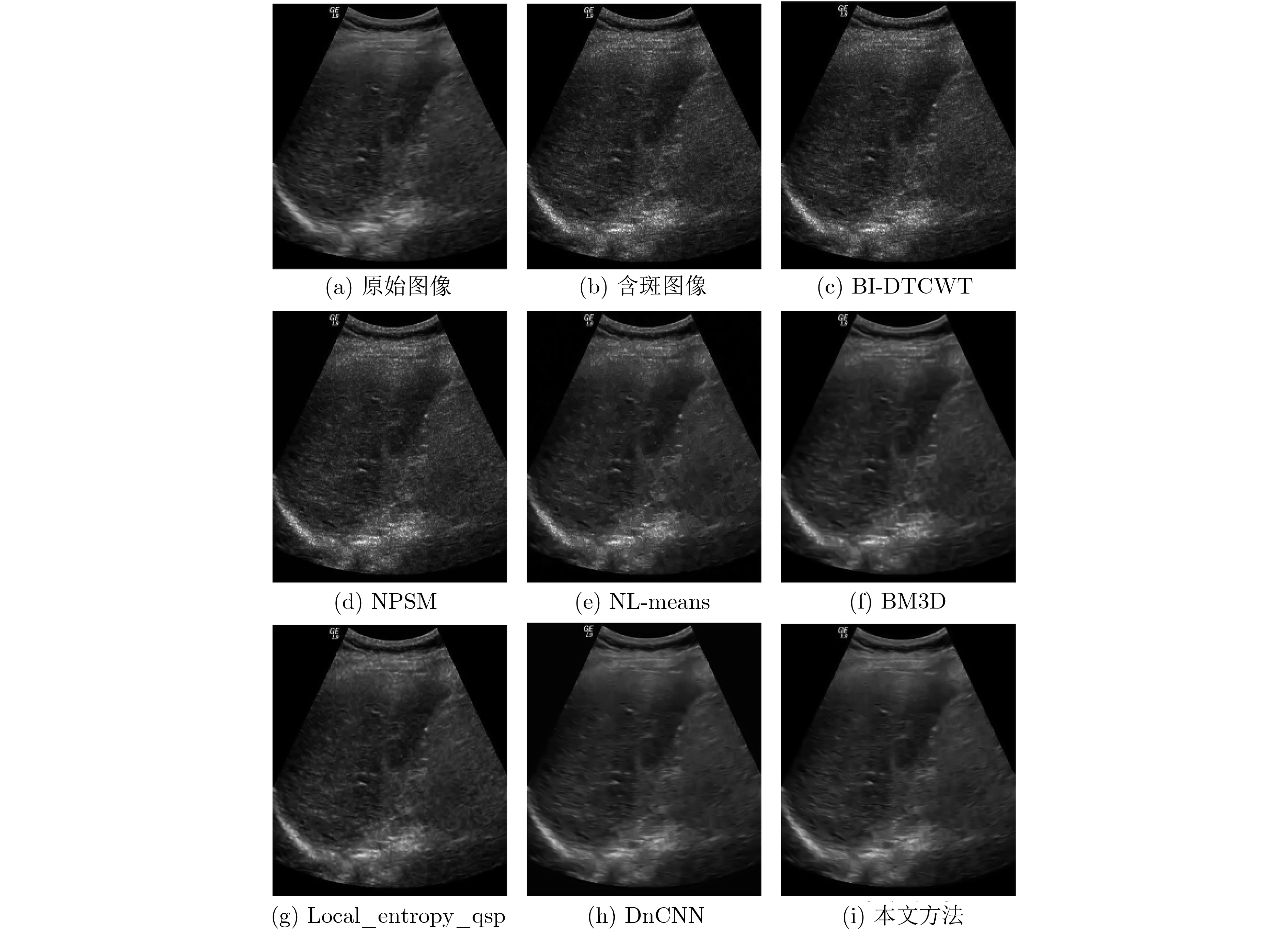
针对传统医学超声图像去斑方法的不足,该文提出一种自适应多曝光融合框架和前馈卷积神经网络模型图像去斑方法。首先,制作超声图像训练数据集;然后,提出一种自适应增强因子的多曝光融合框架,增强图像进行有效特征提取;最后,通过网络训练去斑模型并获得去斑后的图像。实验结果表明,该文较已有的方法,能更有效地滤除医学超声图像中的斑点噪声并更多的保留图像细节。
Abstract:Considering the shortage of traditional medical ultrasound image despeckle methods, an adaptive multi-exposure fusion framework and feedforward convolutional neural network model image despeckle method is proposed. Firstly, an ultrasound image training data set is produced. Then, a multi-exposure fusion framework with adaptive enhancement factors is proposed to enhance the image for effective feature extraction.Finally, a speckle model is trained through the network and a speckle image is obtained. Experimental results show that, compared with the existing methods, this paper can more effectively remove speckle noise in medical ultrasound images and retain more image details.
-
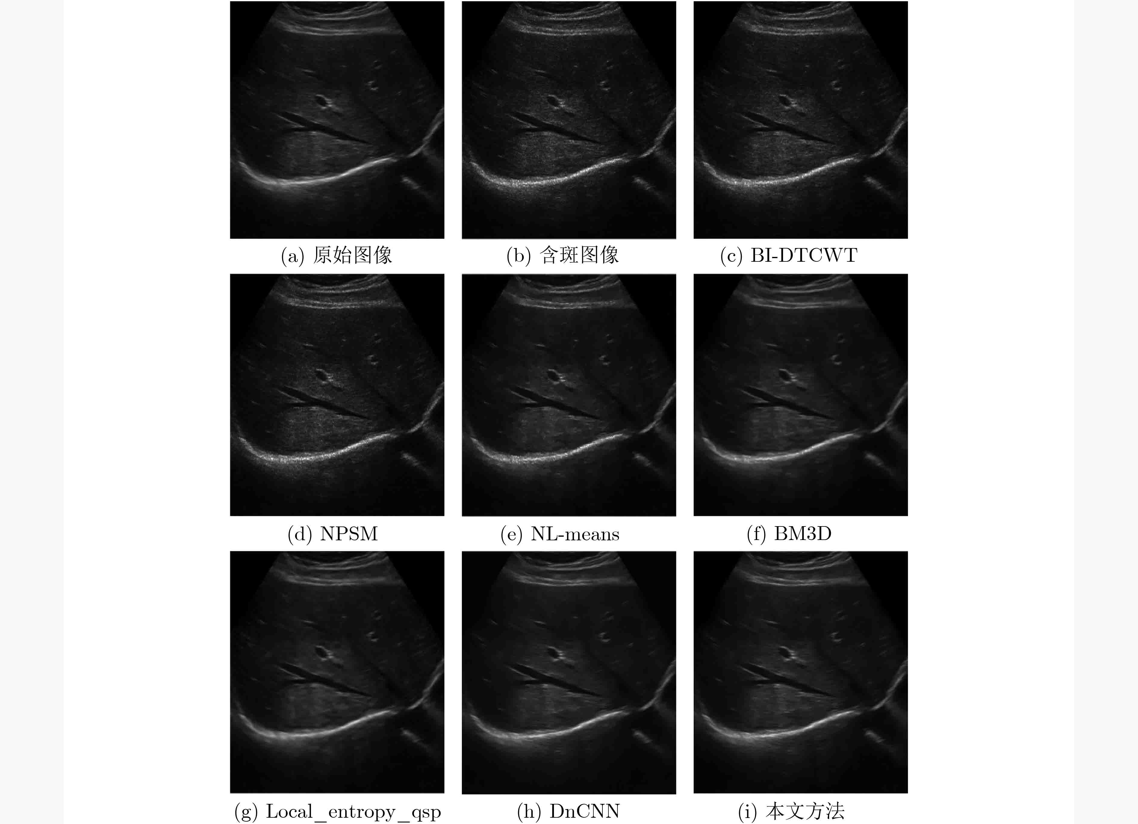
表 1 模拟斑点肝脏超声图像1不同方法PSNR结果(dB)
方法 斑点噪声的标准差σ 0.5 0.6 0.7 0.8 0.9 BI-DTCWT 35.4225 33.8461 32.5368 31.4265 30.3688 NPSM 34.5827 32.9573 31.5366 30.3250 29.2056 NL-means 34.9289 34.1934 33.3554 32.5658 31.6134 BM3D 36.1701 35.7552 35.2485 34.8951 34.2035 Local_entropy_qsp 36.7812 36.1083 35.4363 35.0726 34.5014 DnCNN 35.7701 35.8394 35.8180 35.6769 35.3885 本文方法 36.7203 36.7139 36.6025 36.3568 35.9492  下载: 导出CSV
表 4 模拟斑点肝脏超声图像2不同方法
$\beta $ 结果方法 斑点噪声的标准差σ 0.5 0.6 0.7 0.8 0.9 BI-DTCWT 0.7078 0.6359 0.5612 0.5099 0.4661 NPSM 0.6830 0.6197 0.5479 0.4990 0.4517 NL-means 0.7191 0.6761 0.6037 0.5449 0.4899 BM3D 0.8030 0.7950 0.7826 0.7683 0.7355 Local_entropy_qsp 0.8263 0.8090 0.7787 0.7567 0.7384 DnCNN 0.9286 0.9238 0.9156 0.9029 0.8812 本文方法 0.9394 0.9325 0.9217 0.9653 0.8836
下载: 导出CSV
表 2 模拟斑点肝脏超声图像2不同方法PSNR结果(dB)
方法 斑点噪声的标准差σ 0.5 0.6 0.7 0.8 0.9 BI-DTCWT 31.0477 29.5409 28.0856 27.4342 26.2056 NPSM 31.5374 30.0985 28.6745 27.6699 26.6843 NL-means 32.7360 31.7539 30.4860 29.5105 28.4174 BM3D 33.8786 33.3096 32.5436 32.0199 31.2079 Local_entropy_qsp 34.3157 33.2426 32.1706 31.5329 30.8599 DnCNN 34.9760 35.0382 34.8497 34.3851 33.6562 本文方法 35.9280 35.9170 35.6289 35.0301 34.1677
下载: 导出CSV
表 3 模拟斑点肝脏超声图像1不同方法
$\beta $ 结果方法 斑点噪声的标准差σ 0.5 0.6 0.7 0.8 0.9 BI-DTCWT 0.6416 0.5611 0.4823 0.4291 0.3846 NPSM 0.5972 0.5154 0.4352 0.3817 0.3393 NL-means 0.4522 0.4102 0.3564 0.3262 0.2949 BM3D 0.5969 0.5820 0.5685 0.5477 0.5016 Local_entropy_qsp 0.6540 0.6287 0.5991 0.5842 0.5621 DnCNN 0.7803 0.7726 0.7595 0.7393 0.7106 本文方法 0.8128 0.8011 0.7831 0.7564 0.7208
下载: 导出CSV
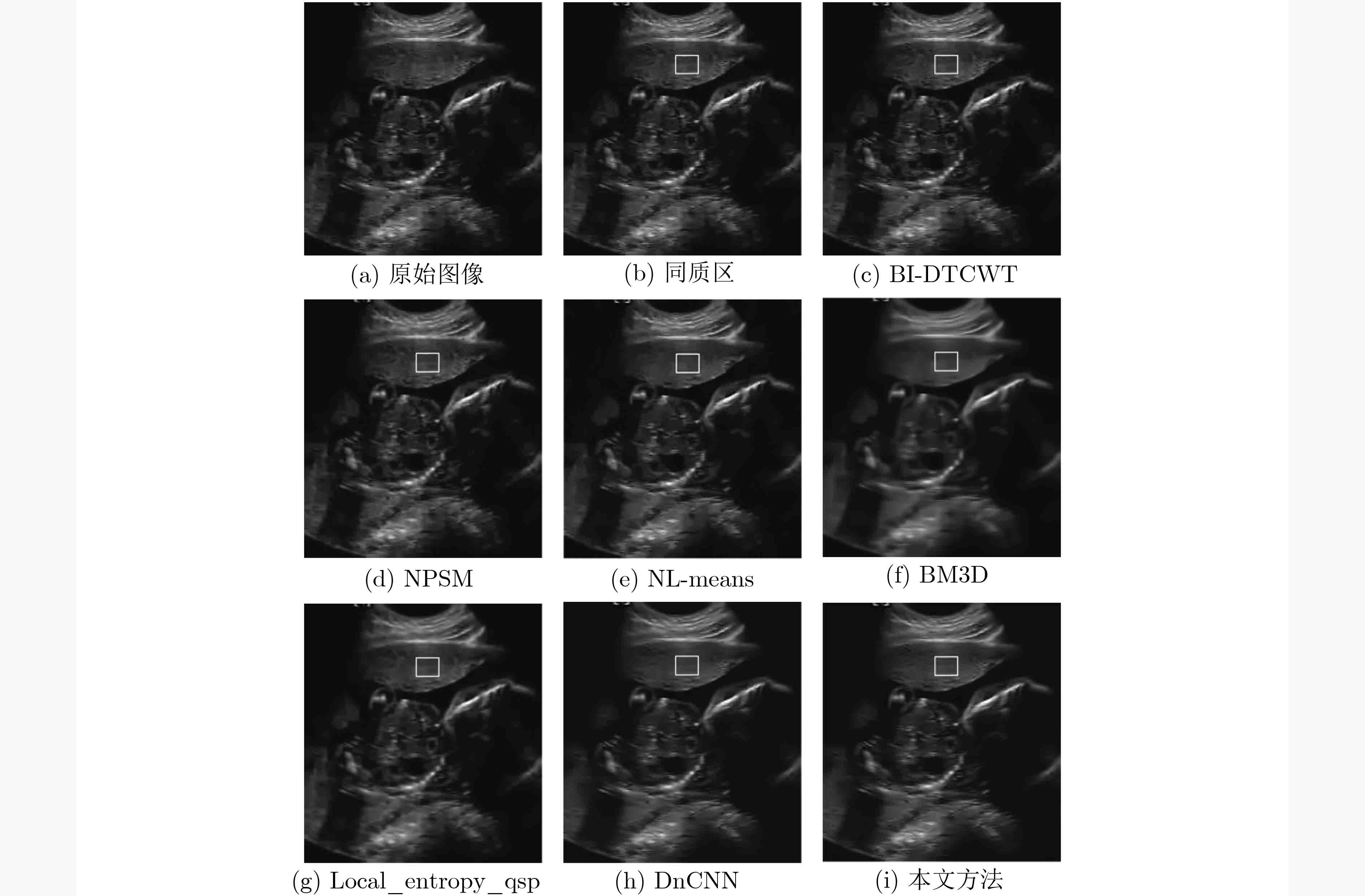
表 5 真实斑点超声图像不同方法ENL结果
方法 ENL等效视数值 BI-DTCWT 61.2209 NPSM 64.6016 NL-means 109.5584 BM3D 93.4877 Local_entropy_qsp 79.1016 DnCNN 132.9184 本文方法 134.3287
下载: 导出CSV
表 6 50张真实斑点超声图像不同方法ENL平均值比较
方法 ENL等效视数值平均值 BI-DTCWT 75.5182 NPSM 75.5941 NL-means 110.6393 BM3D 110.9127 Local_entropy_qsp 93.7911 DnCNN 140.3622 本文方法 147.0689
下载: 导出CSV
-
KANAYAMA Y and YANO M. Ultrasound diagnosis apparatus and ultrasound imaging method[P]. USA Patent, 10231710, 2019. ZHOU Yingyue, ZANG Hongbin, XU Su, et al. An iterative speckle filtering algorithm for ultrasound images based on bayesian nonlocal means filter model[J]. Biomedical Signal Processing and Control, 2019, 48: 104–117. doi: 10.1016/j.bspc.2018.09.011 JOEL T and SIVAKUMAR R. An extensive review on Despeckling of medical ultrasound images using various transformation techniques[J]. Applied Acoustics, 2018, 138: 18–27. doi: 10.1016/j.apacoust.2018.03.023 沈民奋, 陈婷婷, 张琼, 等. 医用超声图像散斑去噪方法综述[J]. 中国医疗器械信息, 2013, 19(3): 17–22. doi: 10.3969/j.issn.1006-6586.2013.03.003SHEN Minfen, CHEN Tingting, ZHANG Qiong, et al. The review of speckle denoising in medical ultrasound imaging[J]. China Medical Device Information, 2013, 19(3): 17–22. doi: 10.3969/j.issn.1006-6586.2013.03.003 KUAN D, SAWCHUK A, STRAND T, et al. Adaptive restoration of images with speckle[J]. IEEE Transactions on Acoustics, Speech, and Signal Processing, 1987, 35(3): 373–383. doi: 10.1109/TASSP.1987.1165131 DABOV K, FOI A, KATKOVNIK V, et al. Image denoising by sparse 3-D transform-domain collaborative filtering[J]. IEEE Transactions on Image Processing, 2007, 16(8): 2080–2095. doi: 10.1109/TIP.2007.901238 肖佳, 张俊华, 梅礼晔. 改进的三维块匹配去噪算法[J]. 计算机科学, 2019, 46(6): 288–294. doi: 10.11896/j.issn.1002-137X.2019.06.043XIAO Jia, ZHANG Junhua, and MEI Liye. Improved block-matching 3D denoising algorithm[J]. Computer Science, 2019, 46(6): 288–294. doi: 10.11896/j.issn.1002-137X.2019.06.043 PERONA P and MALIK J. Scale-space and edge detection using anisotropic diffusion[J]. IEEE Transactions on Pattern Analysis and Machine Intelligence, 1990, 12(7): 629–639. doi: 10.1109/34.56205 YU Yongjian and ACTON S T. Speckle reducing anisotropic diffusion[J]. IEEE Transactions on Image Processing, 2002, 11(11): 1260–1270. doi: 10.1109/TIP.2002.804276 TIAN Jing and CHEN Li. Image despeckling using a non-parametric statistical model of wavelet coefficients[J]. Biomedical Signal Processing and Control, 2011, 6(4): 432–437. doi: 10.1016/j.bspc.2010.11.006 SENDUR L and SELESNICK I W. Bivariate shrinkage functions for wavelet-based denoising exploiting interscale dependency[J]. IEEE Transactions on Signal Processing, 2002, 50(11): 2744–2756. doi: 10.1109/TSP.2002.804091 BURGER H C, SCHULER C J, and HARMELING S. Image denoising: Can plain neural networks compete with BM3D?[C]. 2012 IEEE Conference on Computer Vision and Pattern Recognition, Providence, USA, 2012: 2392–2399. SCHMIDT U and ROTH S. Shrinkage fields for effective image restoration[C]. The IEEE Conference on Computer Vision and Pattern Recognition, Columbus, USA, 2014: 2774–2781. CHEN Yunjin and POCK T. Trainable nonlinear reaction diffusion: A flexible framework for fast and effective image restoration[J]. IEEE Transactions on Pattern Analysis and Machine Intelligence, 2017, 39(6): 1256–1272. doi: 10.1109/TPAMI.2016.2596743 MAO Xiaojiao, SHEN Chunhua, and YANG Yubin. Image restoration using very deep convolutional encoder-decoder networks with symmetric skip connections[C]. Advances in Neural Information Processing Systems, Red Hook, USA, 2016: 2802–2810. ZHANG Kai, ZUO Wangmeng, CHEN Yunjin, et al. Beyond a Gaussian denoiser: Residual learning of deep CNN for image denoising[J]. IEEE Transactions on Image Processing, 2017, 26(7): 3142–3155. doi: 10.1109/TIP.2017.2662206 NADEEM M, HUSSAIN A, and MUNIR A. Fuzzy logic based computational model for speckle noise removal in ultrasound images[J]. Multimedia Tools and Applications, 2019, 78(9): 18531–18548. 吕晓琪, 吴凉, 谷宇, 等. 基于深度卷积神经网络的低剂量CT肺部去噪[J]. 电子与信息学报, 2018, 40(6): 1353–1359. doi: 10.11999/JEIT170769LÜ Xiaoqi, WU Liang, GU Yu, et al. Low dose CT lung denoising model based on deep convolution neural network[J]. Journal of Electronics &Information Technology, 2018, 40(6): 1353–1359. doi: 10.11999/JEIT170769 YING Zhenqiang, LI Ge, REN Yurui, et al. A new image contrast enhancement algorithm using exposure fusion framework[C]. The 17th International Conference on Computer Analysis of Images and Patterns, Ystad, Sweden, 2017: 36–46. ZHENG D, WANG J, and XIAO Z. Image enhancement based on local standard deviation[J]. Journal of Information and Computational Science, 2005, 2(2): 429–437. JIFARA W, JIANG Feng, RHO S, et al. Medical image denoising using convolutional neural network: A residual learning approach[J]. The Journal of Supercomputing, 2019, 75(2): 704–718. doi: 10.1007/s11227-017-2080-0 SIMONYAN K and ZISSERMAN A. Very deep convolutional networks for large-scale image recognition[J]. arXiv, 2014, 1409.1556. VEDALDI A and LENC K. MatconvNet: Convolutional neural networks for MATLAB[C]. The 23rd ACM International Conference on Multimedia, Brisbane, Australia, 2015: 689–692. FU Xiaowei, WANG Yi, CHEN Li, et al. An image despeckling approach using quantum-inspired statistics in dual-tree complex wavelet domain[J]. Biomedical Signal Processing and Control, 2015, 18: 30–35. doi: 10.1016/j.bspc.2014.11.005 BUADES A, COLL B, and MOREL J M. A non-local algorithm for image denoising[C]. 2005 IEEE Computer Society Conference on Computer Vision and Pattern Recognition, San Diego, USA, 2005: 60–65. 付晓薇, 代芸, 陈黎, 等. 基于局部熵的量子衍生医学超声图像去斑[J]. 电子与信息学报, 2015, 37(3): 560–566. doi: 10.11999/JEIT140587FU Xiaowei, DAI Wei, CHEN Li, et al. Quantum-inspired despeckling of medical ultrasound images based on local entropy[J]. Journal of Electronics &Information Technology, 2015, 37(3): 560–566. doi: 10.11999/JEIT140587 PIZURICA A, PHILIPS W, LEMAHIEU I, et al. A versatile wavelet domain noise filtration technique for medical imaging[J]. IEEE Transactions on Medical Imaging, 2003, 22(3): 323–331. doi: 10.1109/TMI.2003.809588 RODTOOK A and MAKHANOV S S. Multi-feature gradient vector flow snakes for adaptive segmentation of the ultrasound images of breast cancer[J]. Journal of Visual Communication and Image Representation, 2013, 24(8): 1414–1430. doi: 10.1016/j.jvcir.2013.09.009 -


计量
- 文章访问数: 4198
- HTML全文浏览量: 1950
- PDF下载量: 180
- 被引次数: 0
 下载:
下载:
