| Citation: | HU Min, ZHOU Xiudong, HUANG Hongcheng, ZHANG Guanghua, TAO Yang. Computed-Tomography Image Segmentation of Cerebral Hemorrhage Based on Improved U-shaped Neural Network[J]. Journal of Electronics & Information Technology, 2022, 44(1): 127-137. doi: 10.11999/JEIT200996

|

| [1] |
谈山峰, 方芳, 陈兵, 等. 脑疝后脑梗塞预后因素分析[J]. 海南医学, 2014, 25(3): 400–402. doi: 10.3969/j.issn.1003-6350.2014.03.0152
TAN Shanfeng, FANG Fang, CHEN Bing, et al. Analysis of prognostic factors of cerebral infarction after cerebral hernia[J]. Hainan Medical Journal, 2014, 25(3): 400–402. doi: 10.3969/j.issn.1003-6350.2014.03.0152
|
| [2] |
SUN Mingjie, HU R, YU Huimin, et al. Intracranial hemorrhage detection by 3D voxel segmentation on brain CT images[C]. 2015 International Conference on Wireless Communications & Signal Processing (WCSP), Nanjing, China, 2015: 1–5. doi: 10.1109/WCSP.2015.7341238.
|
| [3] |
WANG Nian, TONG Fei, TU Yongcheng, et al. Extraction of cerebral hemorrhage and calculation of its volume on CT image using automatic segmentation algorithm[J]. Journal of Physics: Conference Series, 2019, 1187(4): 042088. doi: 10.1088/1742-6596/1187/4/042088
|
| [4] |
BHADAURIA H S, SINGH A, and DEWAL M L. An integrated method for hemorrhage segmentation from brain CT Imaging[J]. Computers & Electrical Engineering, 2013, 39(5): 1527–1536. doi: 10.1016/j.compeleceng.2013.04.010
|
| [5] |
SHAHANGIAN B and POURGHASSEM H. Automatic brain hemorrhage segmentation and classification in CT scan images[C]. 2013 8th Iranian Conference on Machine Vision and Image Processing (MVIP), Zanjan, Iran, 2013: 467–471. doi: 10.1109/IranianMVIP.2013.6780031.
|
| [6] |
KRIZHEVSKY A, SUTSKEVER I, and HINTON G E. ImageNet classification with deep convolutional neural networks[J]. Communications of the ACM, 2017, 60(6): 84–90. doi: 10.1145/3065386
|
| [7] |
WANG Shuxin, CAO Shilei, WEI Dong, et al. LT-Net: Label transfer by learning reversible voxel-wise correspondence for one-shot medical image segmentation[C]. 2020 IEEE/CVF Conference on Computer Vision and Pattern Recognition (CVPR), Seattle, USA, 2020: 9159–9168. doi: 10.1109/CVPR42600.2020.00918.
|
| [8] |
RONNEBERGER O, FISCHER P, and BROX T. U-Net: Convolutional networks for biomedical image segmentation[C]. The 18th International Conference on Medical Image Computing and Computer-Assisted Intervention, Munich, Germany, 2015: 234–241. doi: 10.1007/978-3-319-24574-4_28.
|
| [9] |
彭佳林, 揭萍. 基于序列间先验约束和多视角信息融合的肝脏CT图像分割[J]. 电子与信息学报, 2018, 40(4): 971–978. doi: 10.11999/JEIT170933
PENG Jialin and JIE Ping. Liver segmentation from CT image based on sequential constraint and multi-view information fusion[J]. Journal of Electronics &Information Technology, 2018, 40(4): 971–978. doi: 10.11999/JEIT170933
|
| [10] |
MILLETARI F, NAVAB N, and AHMADI S A. V-Net: Fully convolutional neural networks for volumetric medical image segmentation[C]. 2016 Fourth International Conference on 3D Vision (3DV), Stanford, USA, 2016: 565–571. doi: 10.1109/3DV.2016.79.
|
| [11] |
GUAN S, KHAN A A, SIKDAR S, et al. Fully dense UNet for 2-D sparse photoacoustic tomography artifact removal[J]. IEEE Journal of Biomedical and Health Informatics, 2020, 24(2): 568–576. doi: 10.1109/JBHI.2019.2912935
|
| [12] |
XIAO Xiao, LIAN Shen, LUO Zhiming, et al. Weighted Res-UNet for high-quality retina vessel segmentation[C]. 2018 9th International Conference on Information Technology in Medicine and Education (ITME), Hangzhou, China, 2018: 327–331. doi: 10.1109/ITME.2018.00080.
|
| [13] |
OKTAY O, SCHLEMPER J, LE FOLGOC L, et al. Attention U-Net: Learning where to look for the pancreas[C]. The 1st Conference on Medical Imaging with Deep Learning, Amsterdam, Netherlands, 2018: 1–10.
|
| [14] |
IBTEHAZ N and RAHMAN M S. MultiResUNet: Rethinking the U-Net architecture for multimodal biomedical image segmentation[J]. Neural Networks, 2020, 121: 74–87. doi: 10.1016/j.neunet.2019.08.025
|
| [15] |
ALOM M Z, YAKOPCIC C, TAHA T M, et al. Nuclei segmentation with recurrent residual convolutional neural networks based U-Net (R2U-Net)[C]. NAECON 2018-IEEE National Aerospace and Electronics Conference, Dayton, USA, 2018: 228–233. doi: 10.1109/NAECON.2018.8556686.
|
| [16] |
ZHOU Zongwei, RAHMAN M M, TAJBAKHSH N, et al. UNet++: Redesigning skip connections to exploit multiscale features in image segmentation[J]. IEEE Transactions on Medical Imaging, 2020, 39(6): 1856–1867. doi: 10.1109/TMI.2019.2959609
|
| [17] |
GU Zaiwang, CHENG Jun, FU Huazhu, et al. CE-Net: Context encoder network for 2D medical image segmentation[J]. IEEE Transactions on Medical Imaging, 2019, 38(10): 2281–2292. doi: 10.1109/TMI.2019.2903562
|
| [18] |
FU Jun, LIU Jing, TIAN Haijie, et al. Dual attention network for scene segmentation[C]. 2019 IEEE/CVF Conference on Computer Vision and Pattern Recognition (CVPR), Long Beach, USA, 2019: 3141–3149. doi: 10.1109/CVPR.2019.00326.
|
| [19] |
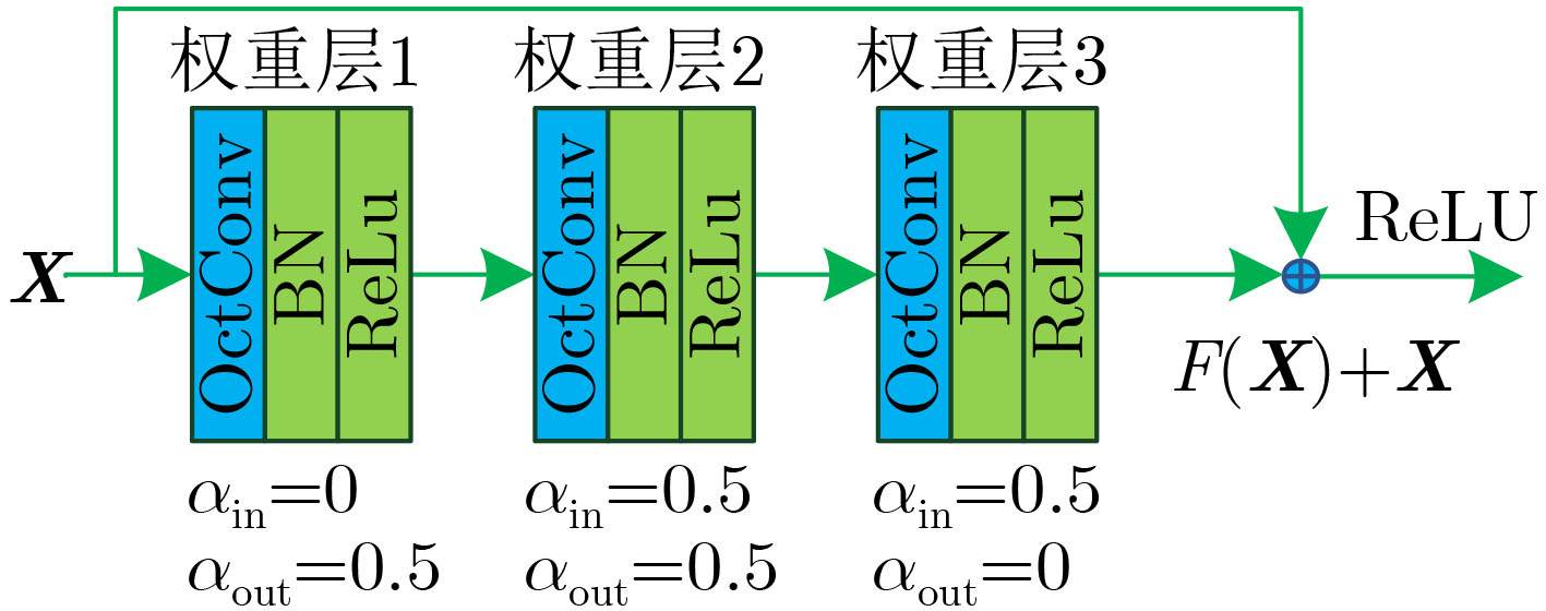
CHEN Yunpeng, FAN Haoqi, XU Bing, et al. Drop an Octave: Reducing spatial redundancy in convolutional neural networks with Octave convolution[C]. 2019 IEEE/CVF International Conference on Computer Vision (ICCV), Seoul, South Korea, 2019: 3434–3443. doi: 10.1109/ICCV.2019.00353.
|

Figures(13) / Tables(6)


 DownLoad: