Advanced Search
Turn off MathJax
Article Contents
YAN Bing, JI Jie, PENG Yuming, NIU Yanxiong, SUN Chang, DING Ming. Design and Quantitative Evaluation of the Fitting Range of a Rigid Helmet for Optically Pumped Magnetometer Magnetoencephalography[J]. Journal of Electronics & Information Technology. doi: 10.11999/JEIT260860
Citation: YAN Bing, JI Jie, PENG Yuming, NIU Yanxiong, SUN Chang, DING Ming. Design and Quantitative Evaluation of the Fitting Range of a Rigid Helmet for Optically Pumped Magnetometer Magnetoencephalography[J]. Journal of Electronics & Information Technology. doi: 10.11999/JEIT260860

Design and Quantitative Evaluation of the Fitting Range of a Rigid Helmet for Optically Pumped Magnetometer Magnetoencephalography

doi: 10.11999/JEIT260860 cstr: 32379.14.JEIT260860
  • Received Date: 2026-06-16
  • Accepted Date: 2026-10-15
  • Rev Recd Date: 2026-10-14
  • Available Online: 2026-10-22
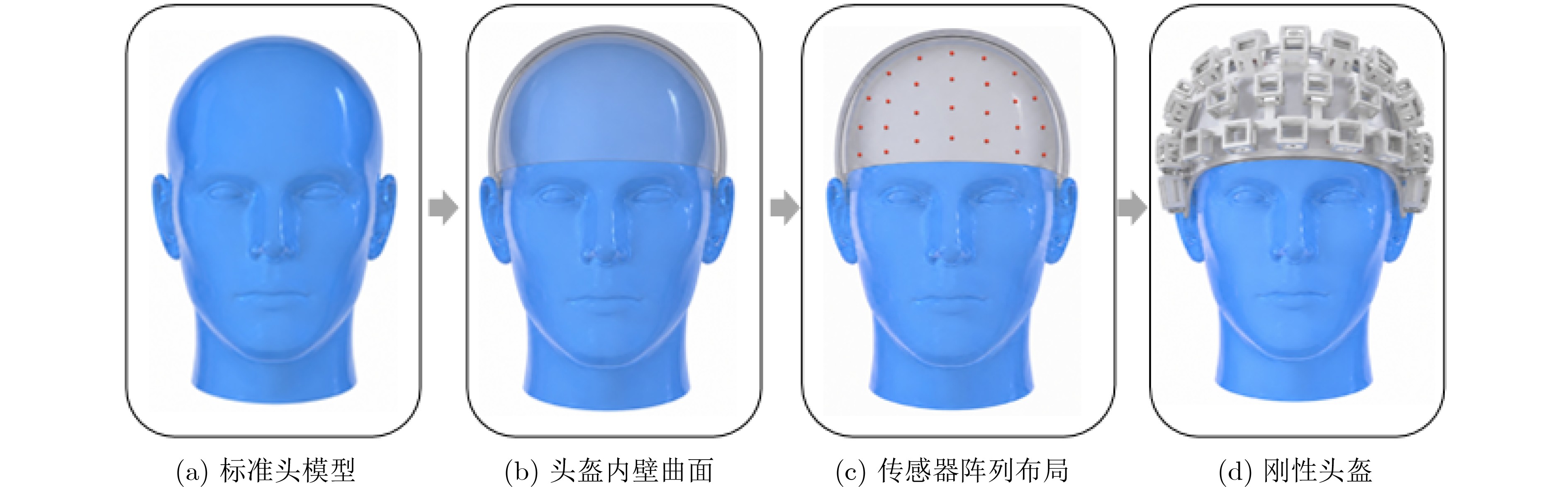
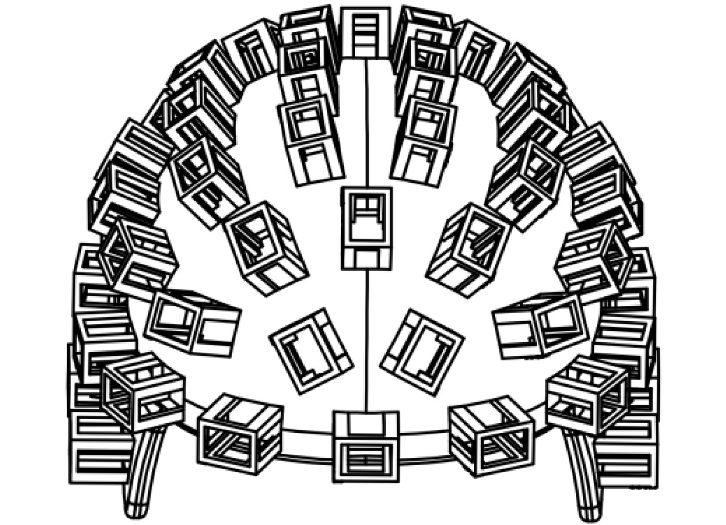
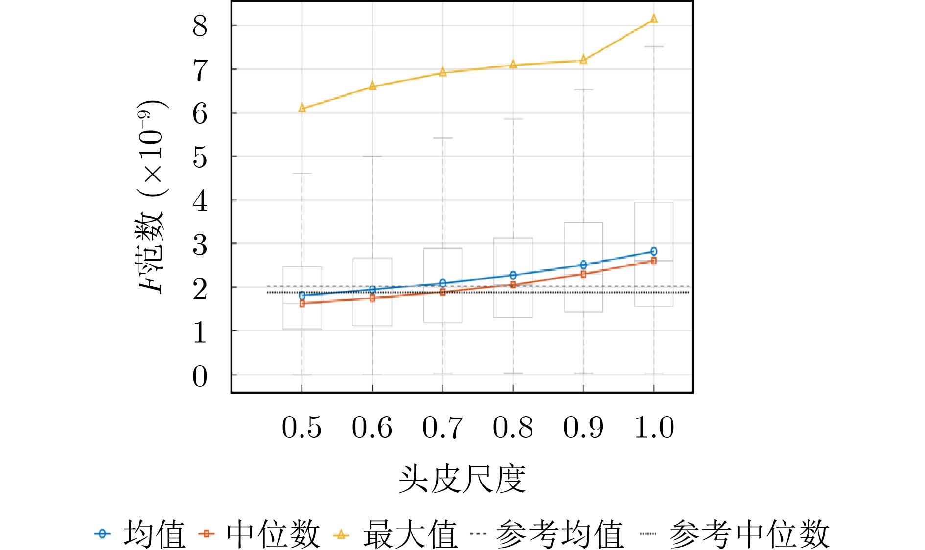
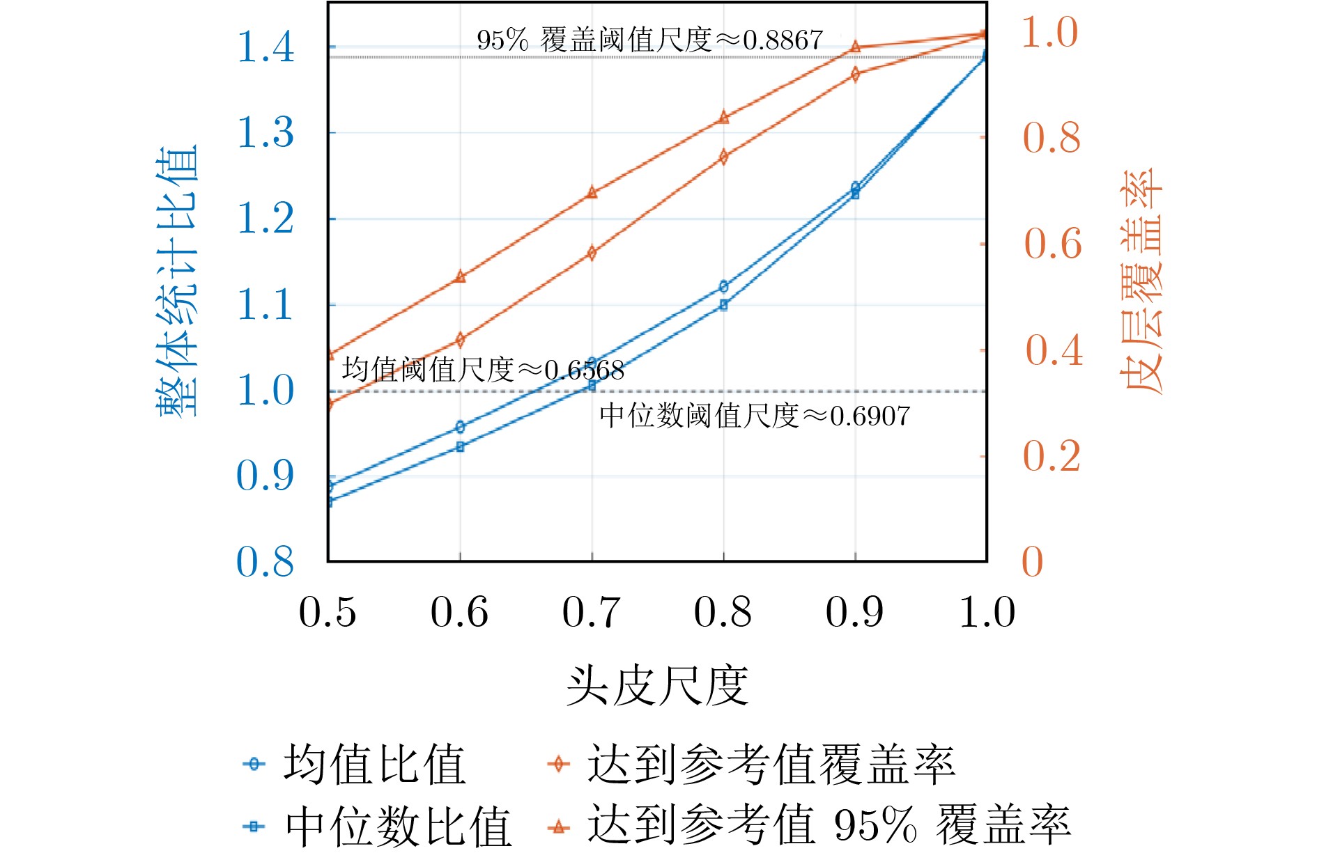
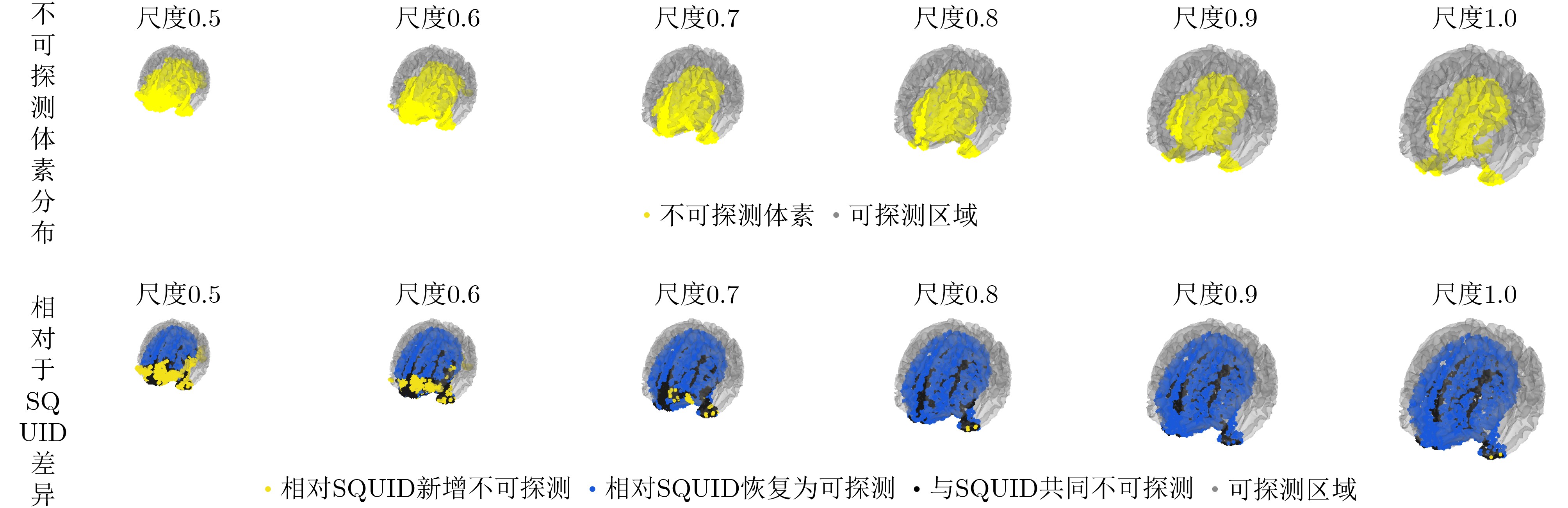
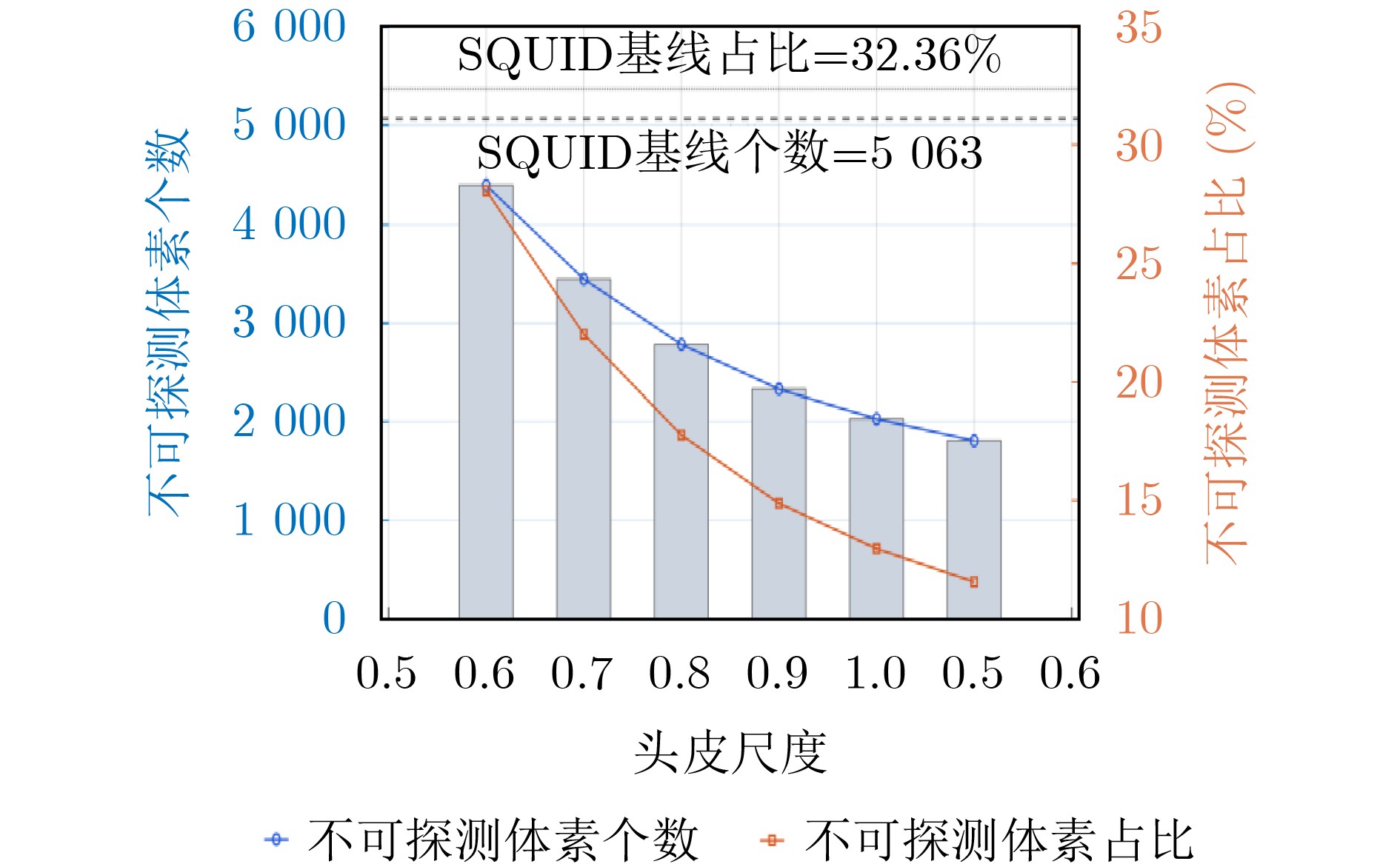
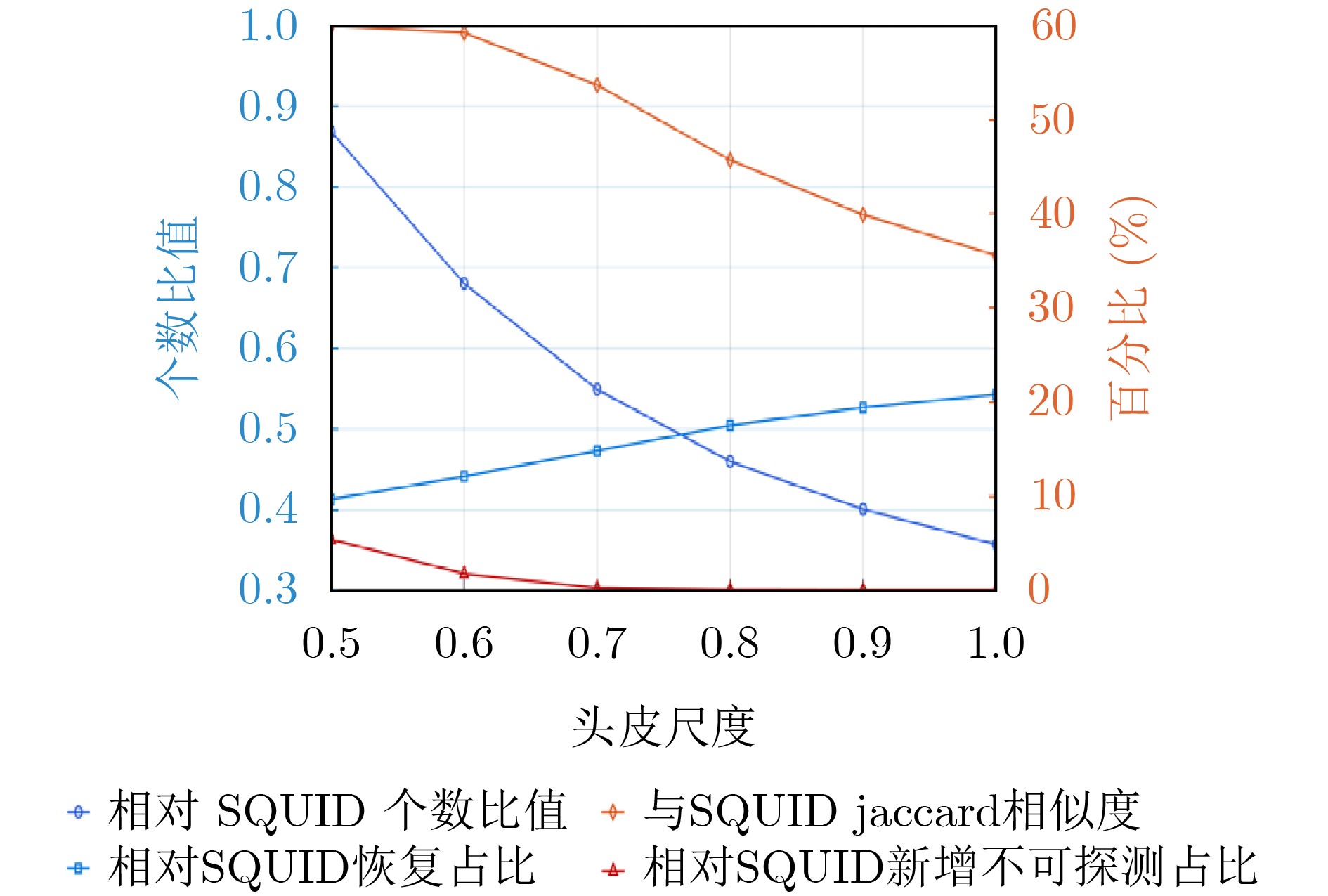
  •   Objective  Magnetoencephalography (MEG) is a non-invasive technique for detecting weak magnetic fields generated by neuronal activity. Compared with conventional superconducting quantum interference device-based MEG systems, optically pumped magnetometer-based magnetoencephalography (OPM-MEG) allows sensors to operate at room temperature and to be placed closer to the scalp, thereby improving signal detection efficiency and supporting wearable system design. In practical OPM-MEG systems, rigid helmets can provide stable sensor positions and improve repeated measurement consistency. However, because head size varies across subjects, a fixed-size rigid helmet may increase the sensor-to-scalp distance for smaller heads, resulting in reduced forward-field strength and limited detectable coverage. Existing OPM-MEG helmet studies have mainly focused on system implementation and experimental validation, whereas quantitative methods for evaluating the size-adaptation range of fixed-size rigid helmets remain insufficient. This study proposes a standard-head-model-based design and multidimensional quantitative evaluation method for an OPM-MEG rigid helmet, aiming to provide guidance for helmet standardization, size planning, and engineering application.  Methods  The ICBM152 standard head model was used as the geometric reference for helmet design. The scalp surface was extracted and truncated according to the brain coverage requirement, and a smooth inner-wall surface was generated. Following the principles of maintaining basic wearing clearance, reducing the sensor-to-cortex distance, and ensuring whole-brain coverage, a 64-channel OPM-MEG sensor array was arranged to cover the frontal, parietal, temporal, and occipital regions. Based on the inner-wall surface and sensor positions, a rigid helmet structure with sensor slots and ear openings was constructed, considering structural stability, 3D-printing feasibility, and wearing comfort. To evaluate the size-adaptation range, the standard head model was defined as the 1.0-scale model, and six head-size models with scale factors of 0.5, 0.6, 0.7, 0.8, 0.9, and 1.0 were generated. During scaling, the helmet and sensor array remained fixed, while the head model was proportionally resized with the top of the head kept unchanged. A 306-channel SQUID-MEG system was used as the reference baseline. The Frobenius norm of the lead field was used to evaluate forward-field detection capability, while undetectable voxels were used to quantify effective detectable coverage. Mean ratio, median ratio, spatial coverage rate, undetectable-voxel ratio, Jaccard similarity, newly undetectable voxels, and recovered detectable voxels were calculated for comparison.  Results and Discussions  The simulation results showed that head-size mismatch had a clear influence on the detection capability of the fixed-size OPM-MEG rigid helmet. As the head scale increased from 0.5 to 1.0, the Frobenius norm over the cortical surface increased continuously, indicating enhanced forward-field strength when cortical sources became closer to the fixed sensor array. The mean Frobenius norm increased from 1.81×10⁻⁹ at the 0.5-scale model to 2.82×10⁻⁹ at the 1.0-scale model, corresponding to 88.9% and 139.0% of the SQUID-MEG reference baseline, respectively. The median ratio showed a similar increase from 87.1% to 139.1%. Based on linear interpolation, the mean and median Frobenius norm ratios reached the SQUID-MEG reference level at head-scale factors of approximately 0.66 and 0.69, respectively. However, when spatial coverage was considered, a stricter threshold was obtained: a head-scale factor of approximately 0.89 was required for 95% of cortical source points to reach at least 95% of the reference detection capability. This indicates that global statistical indicators may reflect the average detection capability, whereas spatial coverage can more sensitively reveal local performance degradation. The undetectable-voxel analysis provided complementary evidence for detectable coverage. As the head scale decreased from 1.0 to 0.5, undetectable regions gradually expanded, mainly in lower and peripheral areas of the region of interest. The number of undetectable voxels increased from 1809 at the 1.0-scale model to 4396 at the 0.5-scale model, corresponding to 11.56% and 28.10% of all voxels, respectively. In contrast, the SQUID-MEG reference system had 5063 undetectable voxels, accounting for 32.36%. Therefore, within the tested range of 0.5–1.0, the designed OPM-MEG rigid helmet maintained fewer undetectable voxels than the SQUID-MEG baseline. Nevertheless, newly undetectable voxels appeared when the head scale decreased to 0.6 and 0.5, indicating local degradation of detectable coverage. These results suggest that the size-adaptation range of a fixed-size rigid helmet is not a single fixed value, but depends on the selected evaluation criterion and application scenario.  Conclusions  This study proposes a standard-head-model-based design and quantitative evaluation method for the size-adaptation range of an OPM-MEG rigid helmet. A 64-channel rigid helmet was designed using the ICBM152 standard head model, and its performance was evaluated under different head-size conditions by combining forward-field strength and detectable-coverage metrics. When the mean and median Frobenius norm ratios were used as overall performance criteria, the lower adaptation limits were approximately 0.66 and 0.69 times the standard head model. When spatial coverage was used as a stricter criterion, the lower limit increased to approximately 0.89. Based on the undetectable-voxel analysis, the helmet maintained better overall detectable coverage than the SQUID-MEG reference within the tested range, while the preferred engineering adaptation limit was approximately 0.7. These findings provide quantitative guidance for OPM-MEG rigid helmet standardization, size planning, applicable population analysis, and engineering application.
  • loading
  • [1]
    COHEN D. Magnetoencephalography: Evidence of magnetic fields produced by alpha-rhythm currents[J]. Science, 1968, 161(3843): 784–786. doi: 10.1126/science.161.3843.784.
    [2]
    COHEN D. Magnetoencephalography: Detection of the brain’s electrical activity with a superconducting magnetometer[J]. Science, 1972, 175(4022): 664–666. doi: 10.1126/science.175.4022.664.
    [3]
    BAILLET S. Magnetoencephalography for brain electrophysiology and imaging[J]. Nature Neuroscience, 2017, 20(3): 327–339. doi: 10.1038/nn.4504.
    [4]
    GROSS J, BAILLET S, BARNES G R, et al. Good practice for conducting and reporting MEG research[J]. NeuroImage, 2013, 65: 349–363. doi: 10.1016/j.neuroimage.2012.10.001.
    [5]
    SHAH V K and WAKAI R T. A compact, high performance atomic magnetometer for biomedical applications[J]. Physics in Medicine and Biology, 2013, 58(22): 8153–8161. doi: 10.1088/0031-9155/58/22/8153.
    [6]
    BOTO E, MEYER S S, SHAH V, et al. A new generation of magnetoencephalography: Room temperature measurements using optically-pumped magnetometers[J]. NeuroImage, 2017, 149: 404–414. doi: 10.1016/j.neuroimage.2017.01.034.
    [7]
    BOTO E, HOLMES N, LEGGETT J, et al. Moving magnetoencephalography towards real-world applications with a wearable system[J]. Nature, 2018, 555(7698): 657–661. doi: 10.1038/nature26147.
    [8]
    BROOKES M J, LEGGETT J, REA M, et al. Magnetoencephalography with optically pumped magnetometers (OPM-MEG): The next generation of functional neuroimaging[J]. Trends in Neurosciences, 2022, 45(8): 621–634. doi: 10.1016/j.tins.2022.05.008.
    [9]
    IIVANAINEN J, STENROOS M, and PARKKONEN L. Measuring MEG closer to the brain: Performance of on-scalp sensor arrays[J]. NeuroImage, 2017, 147: 542–553. doi: 10.1016/j.neuroimage.2016.12.048.
    [10]
    HILL R M, BOTO E, REA M, et al. Multi-channel whole-head OPM-MEG: Helmet design and a comparison with a conventional system[J]. NeuroImage, 2020, 219: 116995. doi: 10.1016/j.neuroimage.2020.116995.
    [11]
    SAFAR K, VANDEWOUW M M, SATO J, et al. Using optically pumped magnetometers to replicate task-related responses in next generation magnetoencephalography[J]. Scientific Reports, 2024, 14(1): 6513. doi: 10.1038/s41598-024-56878-6.
    [12]
    BRICKWEDDE M, ANDERS P, KÜHN A A, et al. Applications of OPM-MEG for translational neuroscience: A perspective[J]. Translational Psychiatry, 2024, 14(1): 341. doi: 10.1038/s41398-024-03047-y.
    [13]
    HILL R M, BOTO E, HOLMES N, et al. A tool for functional brain imaging with lifespan compliance[J]. Nature Communications, 2019, 10(1): 4785. doi: 10.1038/s41467-019-12486-x.
    [14]
    ALEM O, HUGHES K J, BUARD I, et al. An integrated full-head OPM-MEG system based on 128 zero-field sensors[J]. Frontiers in Neuroscience, 2023, 17: 1190310. doi: 10.3389/fnins.2023.1190310.
    [15]
    SCHOFIELD H, HILL R M, FEYS O, et al. A novel, robust, and portable platform for magnetoencephalography using optically-pumped magnetometers[J]. Imaging Neuroscience, 2024, 2: imag-2–00283. doi: 10.1162/imag_a_00283.
    [16]
    HILL R M, REINA RIVERO G, TYLER A J, et al. Determining sensor geometry and gain in a wearable MEG system[J]. Imaging Neuroscience, 2025, 3: imag_a_00535. doi: 10.1162/imag_a_00535.
    [17]
    SCHOFIELD H, HILL R M, RIER L, et al. Towards a 384-channel magnetoencephalography system based on optically pumped magnetometers[J]. Imaging Neuroscience, 2025, 3: IMAG. a. 1042. doi: 10.1162/IMAG.a.1042.
    [18]
    BELTRACHINI L, VON ELLENRIEDER N, EICHARDT R, et al. Optimal design of on‐scalp electromagnetic sensor arrays for brain source localisation[J]. Human Brain Mapping, 2021, 42(15): 4869–4879. doi: 10.1002/hbm.25586.
    [19]
    TIERNEY T M, MELLOR S, O’NEILL G C, et al. Pragmatic spatial sampling for wearable MEG arrays[J]. Scientific Reports, 2020, 10(1): 21609. doi: 10.1038/s41598-020-77589-8.
    [20]
    TAKEDA Y, GOMI T, UMEBAYASHI R, et al. Sensor array design of optically pumped magnetometers for accurately estimating source currents[J]. NeuroImage, 2023, 277: 120257. doi: 10.1016/j.neuroimage.2023.120257.
    [21]
    SCHOFFELEN J M, CHEUNG T, KNAPPE S, et al. Optimal configuration of on-scalp OPMs with fixed channel counts[J]. Imaging Neuroscience, 2025, 3: IMAG. a. 22. doi: 10.1162/IMAG.a.22.
    [22]
    MARHL U, HREN R, SANDER T, et al. Optimization of OPM-MEG layouts with a limited number of sensors[J]. Sensors, 2025, 25(9): 2706. doi: 10.3390/s25092706.
    [23]
    FONOV V, EVANS A, MCKINSTRY R, et al. Unbiased nonlinear average age-appropriate brain templates from birth to adulthood[J]. NeuroImage, 2009, 47(S1): S102. doi: 10.1016/S1053-8119(09)70884-5.
    [24]
    FONOV V, EVANS A C, BOTTERON K, et al. Unbiased average age-appropriate atlases for pediatric studies[J]. NeuroImage, 2011, 54(1): 313–327. doi: 10.1016/j.neuroimage.2010.07.033.
    [25]
    YAN Bing, PENG Yuming, ZHANG Yixiang, et al. From simulation to clinic: Assessing the required channel count for effective clinical use of OPM-MEG systems[J]. NeuroImage, 2025, 314: 121262. doi: 10.1016/j.neuroimage.2025.121262.
    [26]
    YAN Bing, JI Jie, PENG Yuming, et al. A Virtual MEG Helmet approach for improving OPM-MEG performance in rigid helmets: Simulations and phantom experiment validation[J]. Biocybernetics and Biomedical Engineering, 2026, 46(2): 455–469. doi: 10.1016/j.bbe.2026.05.001.
  • 加载中

Catalog

    通讯作者: 陈斌, bchen63@163.com
    • 1. 

      沈阳化工大学材料科学与工程学院 沈阳 110142

    1. 本站搜索
    2. 百度学术搜索
    3. 万方数据库搜索
    4. CNKI搜索

    Figures(8)

    Article Metrics

    Article views (75) PDF downloads(9) Cited by()
    Proportional views
    Related

    /

    DownLoad:  Full-Size Img  PowerPoint
    Return
    Return