Advanced Search
Turn off MathJax
Article Contents
YANG Yushi, YAN Jiaxuan, XIE Yi, QIU Lijia, HUANG Danfei. A Lightweight Spatial−Spectral Dual−Branch Transformer Network for Classifying Polarized White Blood Cell Hyperspectral Images[J]. Journal of Electronics & Information Technology. doi: 10.11999/JEIT260124
Citation: YANG Yushi, YAN Jiaxuan, XIE Yi, QIU Lijia, HUANG Danfei. A Lightweight Spatial−Spectral Dual−Branch Transformer Network for Classifying Polarized White Blood Cell Hyperspectral Images[J]. Journal of Electronics & Information Technology. doi: 10.11999/JEIT260124

A Lightweight Spatial−Spectral Dual−Branch Transformer Network for Classifying Polarized White Blood Cell Hyperspectral Images

doi: 10.11999/JEIT260124 cstr: 32379.14.JEIT260124
Funds:  National Natural Science Foundation of China (62105245)
  • Received Date: 2026-01-30
  • Accepted Date: 2026-06-29
  • Rev Recd Date: 2026-06-29
  • Available Online: 2026-07-08
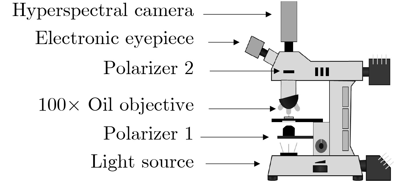
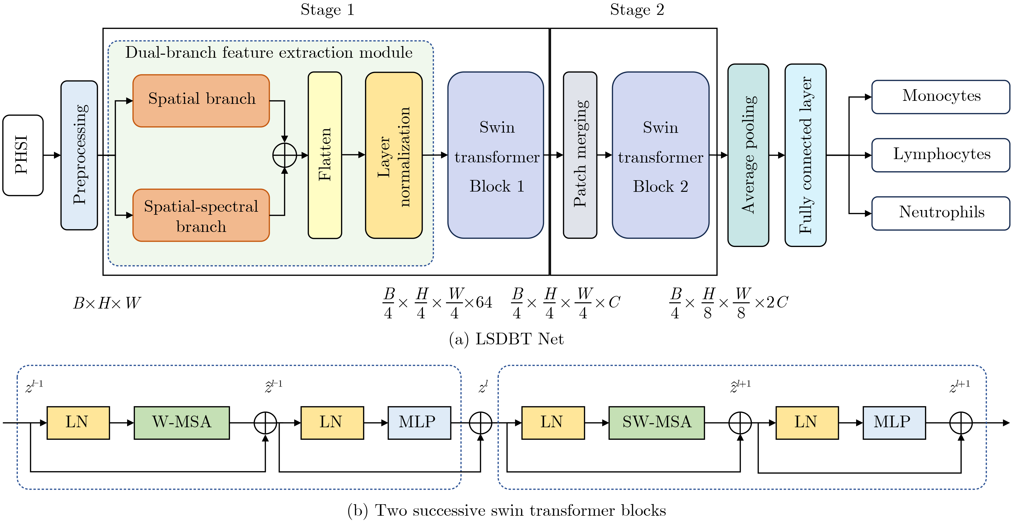
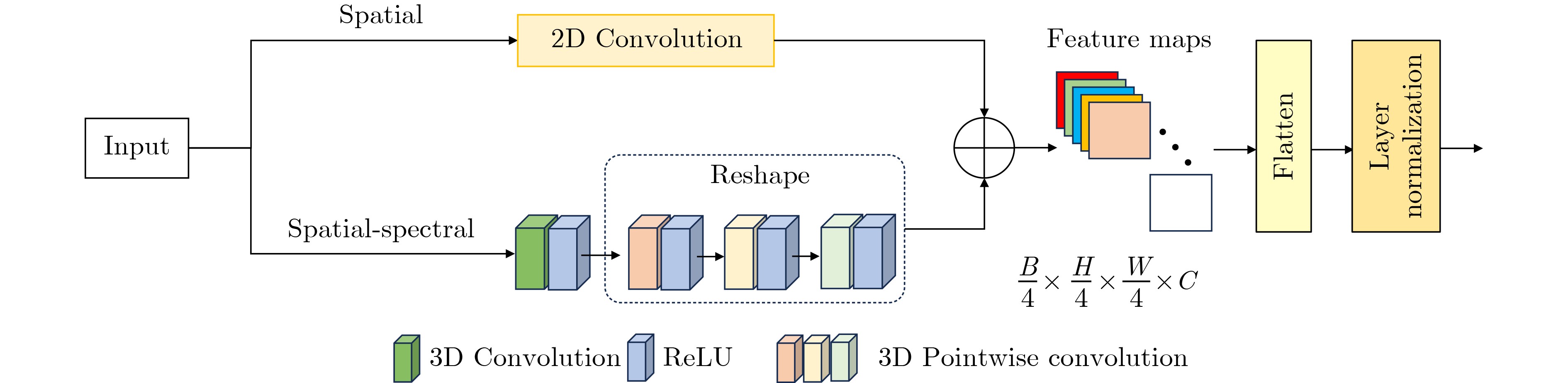
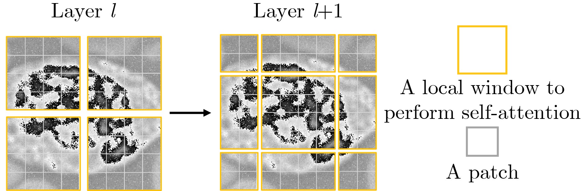
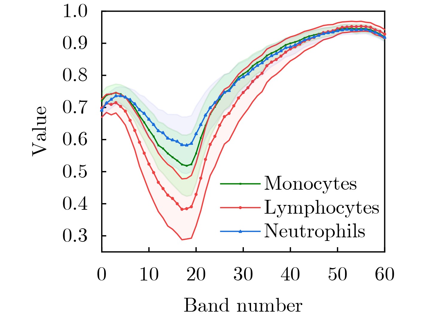
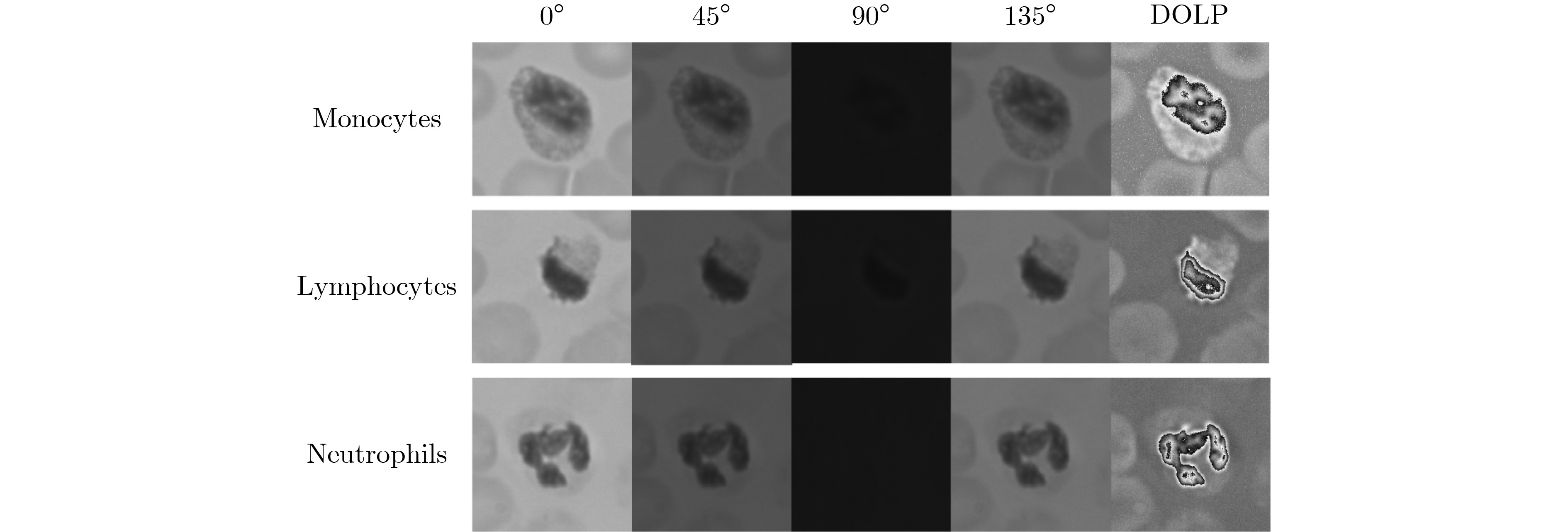
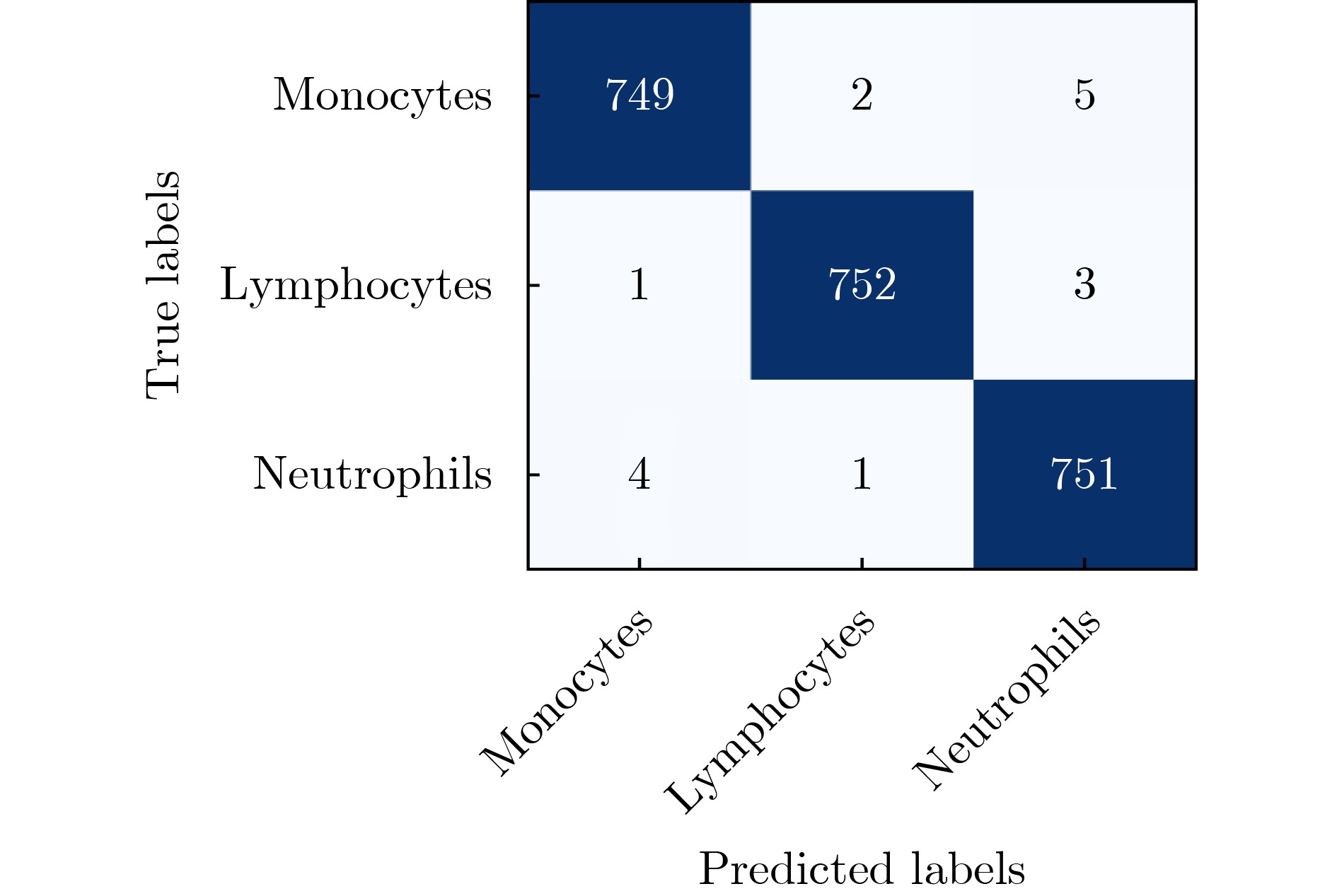
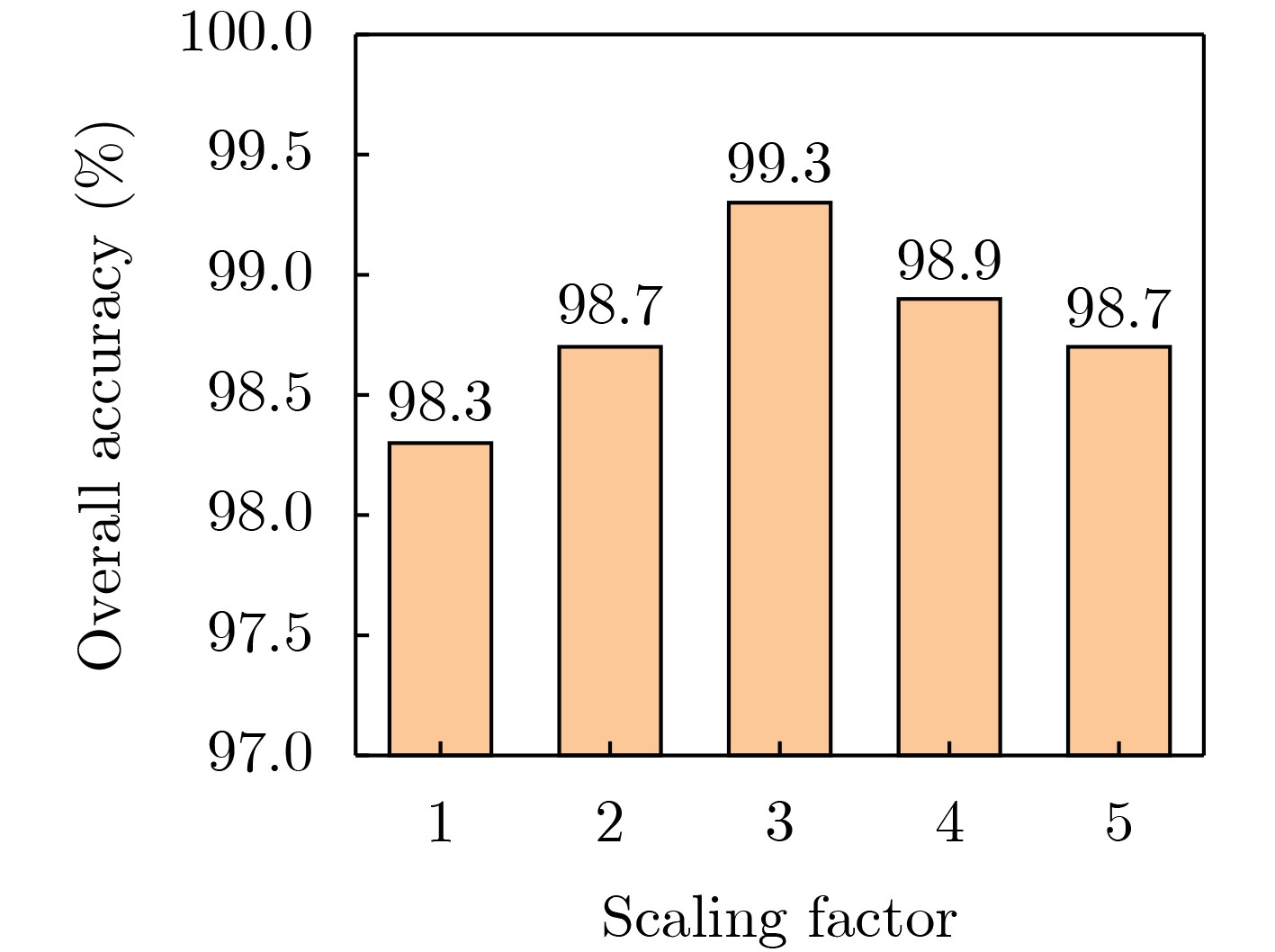
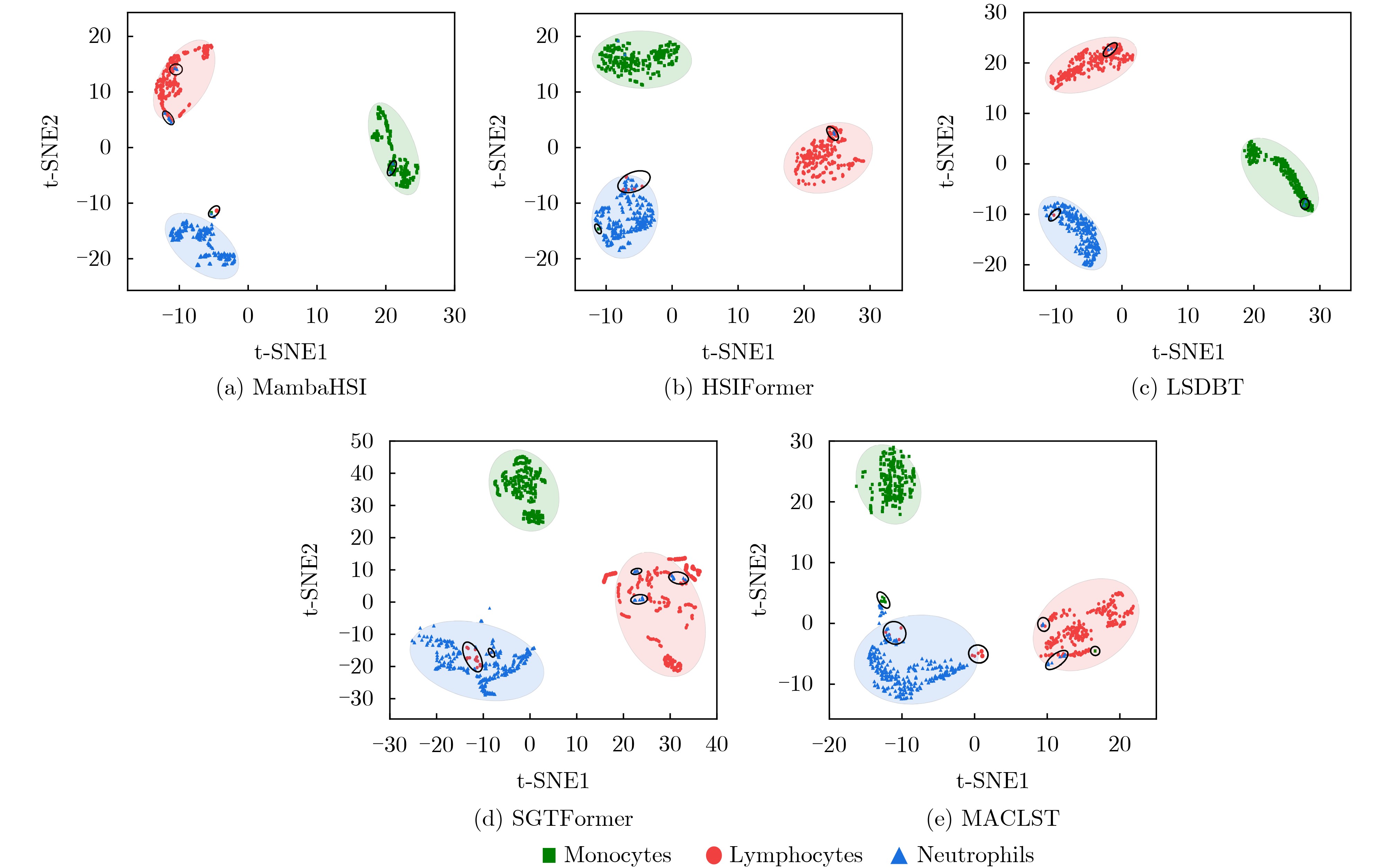
  •   Objective  The classification and morphology of white blood cells (WBC) are pivotal in routine blood analysis, with their precise categorization holding significant importance for disease diagnosis and health assessment. Current WBC classification primarily relies on hematology analyzers and manual microscopy. The former cannot capture cellular images, limiting accuracy when cellular characteristics are abnormal; the latter is constrained by the diagnostician's efficiency and is prone to introducing human error. To enhance the objectivity and efficiency of white blood cell classification, automated analysis and classification methods integrating deep learning and computer vision technologies have emerged as a significant research focus. However, existing methods based on stained images are susceptible to variations in staining conditions and imaging techniques, while traditional hyperspectral imaging remains limited in distinguishing white blood cell subtypes with similar morphological and spectral characteristics. To address this, this study proposes a research approach combining polarized hyperspectral imaging (PHSI) technology with deep learning. It validates the reliability of polarized hyperspectral imaging in white blood cell classification tasks, enabling precise identification of white blood cells and providing an efficient and reliable technical solution for clinical diagnostic support.  Methods  This study first established a polarized hyperspectral microscopic imaging system. By controlling the polarizer at different angles, multiple polarized images were acquired. Based on Stokes vector theory, polarization parameters were calculated to obtain a multidimensional data cube containing both light intensity and polarization state information. Furthermore, linear polarization degree images were computed, thereby constructing a polarized hyperspectral image dataset of white blood cells. Given the multidimensional characteristics of PHSI data, a lightweight spatial−spectral dual−branch Transformer network (LSDBT) is proposed. Preprocessed data are first fed into a dual−branch feature extraction module, which separately extracts local spatial features and spatial−spectral features. An adaptive scaling factor is introduced to fuse these two feature types, thereby fully capturing the multidimensional information inherent in PHSI. The network backbone employs a lightweight Swin Transformer, streamlining the feature learning process to achieve effective global modelling while reducing computational complexity. The model terminates with a classification output via global pooling and a fully connected layer. Its performance in white blood cell classification is systematically evaluated using core metrics: overall accuracy, precision, recall, and F1 score. The network's efficacy in classifying polarized hyperspectral white blood cell images is validated through designed ablation studies, comparative experiments, and feature visualization.  Results and Discussions  The visualization results of DOLP spectral lines and PHSI for white blood cells (Figure 5, Figure 6) reflect the selective absorption and scattering of light by the internal components of monocytes, lymphocytes, and neutrophils. It is evident that the polarization characteristics differ significantly across cell types, and polarized hyperspectral imaging effectively enhances the image contrast of white blood cells, providing polarization information in the spatial dimension that distinguishes it from traditional intensity data. Through experiments with LSDBT, the network achieved an overall accuracy of 99.29% on the test set, with balanced and relatively superior evaluation metrics across all categories (Table 1). This study experimentally investigated the scaling factor within the module, which adjusts the output feature intensity of the spatial−spectral branch. Results (Fig. 8) indicate that model performance initially increases then stabilizes with rising scaling factors, suggesting that appropriately increasing the weight of spatial−spectral features enhances classification performance. The scaling factor was ultimately set to 3. Ablation experiments (Tables 2, Table 3) confirmed the significant contribution of the dual−branch feature extraction module to performance enhancement. Although the lightweight design incurred minor losses, it substantially reduced computational complexity and model parameter count. Compared to hyperspectral imaging, PHSI data demonstrated higher accuracy across all classifiers, proving that polarization information provides unique physical characteristics that enhance cell distinguishability (Table 4). When benchmarked against existing state−of−the−art models, LSDBT achieved optimal performance across multiple metrics (Table 5). Furthermore, t−SNE results (Figure 9) reveal that the features extracted by this network exhibit high clustering and distinct inter−class separation. The computational costs of different models were compared (Table 6), and although LSDBT does not have the lowest computational cost, it achieves the best classification performance while maintaining a balance between model size and computational efficiency.  Conclusions  This study proposes a lightweight dual−branch Transformer network for polarized hyperspectral white blood cell classification, pioneering the integration of polarized hyperspectral imaging with deep learning for white blood cell categorization. Comparative analysis with existing hyperspectral datasets validates the superior performance of this technique in white blood cell classification tasks. In terms of classification model design, the proposed LSDBT network integrates spatial and spectral information through a dual−branch feature extraction module, achieving efficient modelling and classification via lightweight operations. Results demonstrate that this design achieves high feature discriminative power and distinctiveness while reducing computational complexity, yielding excellent performance in polarized hyperspectral white blood cell image classification. This not only provides a high−precision, lightweight method for automated white blood cell classification but also offers further support for the application of polarized hyperspectral imaging technology in cellular microscopic analysis and clinical auxiliary diagnosis.
  • loading
  • [1]
    林宇晖. 基于人工智能的血常规分析技术在临床应用中的研究进展[J]. 中国冶金工业医学杂志, 2025, 42(4): 384–385. doi: 10.13586/j.cnki.yjyx1984.2025.04.066.

    LIN Yuhui. Research progress on the clinical application of artificial intelligence−based complete blood count analysis technology[J]. Chinese Medical Journal of Metallurgical Industry, 2025, 42(4): 384–385.(查阅网上资料, 未找到本条文献英文翻译, 请确认) doi: 10.13586/j.cnki.yjyx1984.2025.04.066.
    [2]
    阮受杰. 比较全自动血液分析仪与血涂片镜检在血常规检验中的准确性差异[J]. 中国医疗器械信息, 2025, 31(12): 88–90. doi: 10.15971/j.cnki.cmdi.2025.12.018.

    RUAN Shoujie. Comparison of accuracy difference between automatic blood analyzer and blood smear microscopic examination in blood routine test[J]. China Medical Device Information, 2025, 31(12): 88–90. doi: 10.15971/j.cnki.cmdi.2025.12.018.
    [3]
    温晶. 全自动血细胞分析仪联合血涂片细胞形态学在血常规检验中的应用效果[J]. 中国医疗器械信息, 2025, 31(12): 91–93. doi: 10.15971/j.cnki.cmdi.2025.12.017.

    WEN Jing. Application effect of automatic hematology analyzer combined with blood smear cell morphology in routine blood tests[J]. China Medical Device Information, 2025, 31(12): 91–93. doi: 10.15971/j.cnki.cmdi.2025.12.017.
    [4]
    ASGHAR R, KUMAR S, SHAUKAT A, et al. Classification of white blood cells (leucocytes) from blood smear imagery using machine and deep learning models: A global scoping review[J]. PLoS One, 2024, 19(6): e0292026. doi: 10.1371/journal.pone.0292026.
    [5]
    WIBAWA M S. A comparison study between deep learning and conventional machine learning on white blood cells classification[C]. Proceedings of 2018 International Conference on Orange Technologies (ICOT), Nusa Dua, Indonesia, 2018. doi: 10.1109/ICOT.2018.8705892.
    [6]
    蔡轶珩, 谭美伶, 潘建军, 等. 基于多尺度非对称密集网络的高光谱图像分类[J]. 电子与信息学报, 2024, 46(4): 1448–1457. doi: 10.11999/JEIT230651.

    CAI Yiheng, TAN Meiling, PAN Jianjun, et al. Hyperspectral image classification based on multi−scale asymmetric dense network[J]. Journal of Electronics & Information Technology, 2024, 46(4): 1448–1457. doi: 10.11999/JEIT230651.
    [7]
    XIE Yi, HUANG Danfei, YAN Jiaxuan, et al. Integrated laser speckle contrast and multispectral imaging for in vivo multimodal assessment of small intestinal viability[J]. Journal of Photochemistry and Photobiology B: Biology, 2025, 272: 113264. doi: 10.1016/j.jphotobiol.2025.113264.
    [8]
    LI Yapeng, LUO Yong, ZHANG Lefei, et al. MambaHSI: Spatial-spectral mamba for hyperspectral image classification[J]. IEEE Transactions on Geoscience and Remote Sensing, 2024, 62: 5524216. doi: 10.1109/tgrs.2024.3430985.
    [9]
    廖帝灵, 赖涛, 黄海风, 等. LightMamba: 一种轻量级Mamba用于高光谱图形和激光雷达数据联合分类网络[J]. 电子与信息学报, 2025, 47(12): 4937–4947. doi: 10.11999/JEIT250981.

    LIAO Diling, LAI Tao, HUANG Haifeng, et al. LightMamba: A lightweight mamba network for the joint classification of HSI and LiDAR data[J]. Journal of Electronics & Information Technology, 2025, 47(12): 4937–4947. doi: 10.11999/JEIT250981.
    [10]
    DUAN Puhong, JIN Shiyu, LU Xiaotian, et al. Domain-adaptive mamba for cross-scene hyperspectral image classification[J]. IEEE Transactions on Image Processing, 2026, 35: 1206–1217. doi: 10.1109/TIP.2026.3657209.
    [11]
    MAHDAVIPOUR Z, ALROMEMA N, KHADER A, et al. TGDHTL: Hyperspectral image classification via transformer-graph convolutional network-diffusion with hybrid domain adaptation[J]. Remote Sensing, 2026, 18(2): 189. doi: 10.3390/rs18020189.
    [12]
    FAROOQUE G, LIU Qichao, SARGANO A B, et al. Swin transformer with multiscale 3D atrous convolution for hyperspectral image classification[J]. Engineering Applications of Artificial Intelligence, 2023, 126: 107070. doi: 10.1016/j.engappai.2023.107070.
    [13]
    ALKHATIB M Q and JAMALI A. HSIFormer: An efficient vision transformer framework for enhanced hyperspectral image classification using local window attention[C]. Proceedings of the 14th Workshop on Hyperspectral Imaging and Signal Processing: Evolution in Remote Sensing (WHISPERS), Helsinki, Finland, 2024: 1–5. doi: 10.1109/WHISPERS65427.2024.10876533.
    [14]
    ZHOU Ximing, MA Ling, HALICEK M, et al. Development of a new polarized hyperspectral imaging microscope[C]. Proceedings of SPIE 11213, Imaging, Therapeutics, and Advanced Technology in Head and Neck Surgery and Otolaryngology, San Francisco, USA, 2020: 1121308. doi: 10.1117/12.2549676.
    [15]
    SONG Yuan, HUANG Danfei, WANG Tianyi, et al. Polarization imaging method for detection of lung cancer based on symmetrical single LCVR[J]. Journal of Biophotonics, 2025, 18(3): e202400470. doi: 10.1002/jbio.202400470.
    [16]
    WOESSNER A E, MCGEE J D, JONES J D, et al. Characterizing differences in the collagen fiber organization of skin wounds using quantitative polarized light imaging[J]. Wound Repair and Regeneration, 2019, 27(6): 711–714. doi: 10.1111/wrr.12758.
    [17]
    ZHOU Ximing, MA Ling, MUBARAK H K, et al. Polarized hyperspectral microscopic imaging system for enhancing the visualization of collagen fibers and head and neck squamous cell carcinoma[J]. Journal of Biomedical Optics, 2024, 29(1): 016005. doi: 10.1117/1.JBO.29.1.016005.
    [18]
    GHASSEMI P, TRAVIS T E, MOFFATT L T, et al. A polarized multispectral imaging system for quantitative assessment of hypertrophic scars[J]. Biomedical Optics Express, 2014, 5(10): 3337–3354. doi: 10.1364/BOE.5.003337.
    [19]
    DREMIN V, MARCINKEVICS Z, ZHEREBTSOV E, et al. Skin complications of diabetes mellitus revealed by polarized hyperspectral imaging and machine learning[J]. IEEE Transactions on Medical Imaging, 2021, 40(4): 1207–1216. doi: 10.1109/TMI.2021.3049591.
    [20]
    ZHOU Ximing, MUBARAK H K, MA Ling, et al. Polarized hyperspectral microscopic imaging for white blood cells on Wright−stained blood smear slides[C]. Proceedings of SPIE 12382, Polarized Light and Optical Angular Momentum for Biomedical Diagnostics, San Francisco, USA, 2023: 1238207. doi: 10.1117/12.2655708.
    [21]
    LIU Ze, LIN Yutong, CAO Yue, et al. Swin transformer: Hierarchical vision transformer using shifted windows[C]. Proceedings of 2021 IEEE/CVF International Conference on Computer Vision (ICCV), Montreal, Canada, 2021: 9992–10022. doi: 10.1109/ICCV48922.2021.00986.
    [22]
    DENG Xiangyu, PAN Lihao, and DANG Zhiyan. SGTformer: Improved shifted window transformer network for white blood cell subtype classification[J]. Journal of Electronic Imaging, 2024, 33(4): 043057. doi: 10.1117/1.JEI.33.4.043057.
    [23]
    FAROOQUE G, LIU Qichao, SARGANO A B, et al. Swin transformer with multiscale 3D atrous convolution for hyperspectral image classification[J]. Engineering Applications of Artificial Intelligence, 2023, 126: 107070.(查阅网上资料, 本条文献和第12条文献重复, 请核对) doi: 10.1016/j.engappai.2023.107070.
  • 加载中

Catalog

    通讯作者: 陈斌, bchen63@163.com
    • 1. 

      沈阳化工大学材料科学与工程学院 沈阳 110142

    1. 本站搜索
    2. 百度学术搜索
    3. 万方数据库搜索
    4. CNKI搜索

    Figures(9)  / Tables(6)

    Article Metrics

    Article views (116) PDF downloads(9) Cited by()
    Proportional views
    Related

    /

    DownLoad:  Full-Size Img  PowerPoint
    Return
    Return