




Reynolds Decomposition Motion-Guided Texture Learning for Scarred Myocardium Phenotyping
-
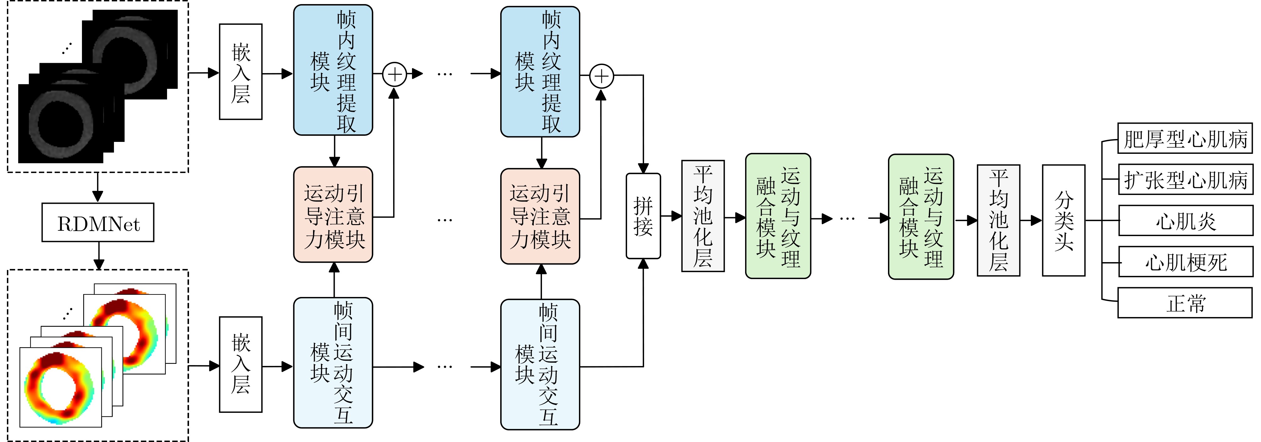
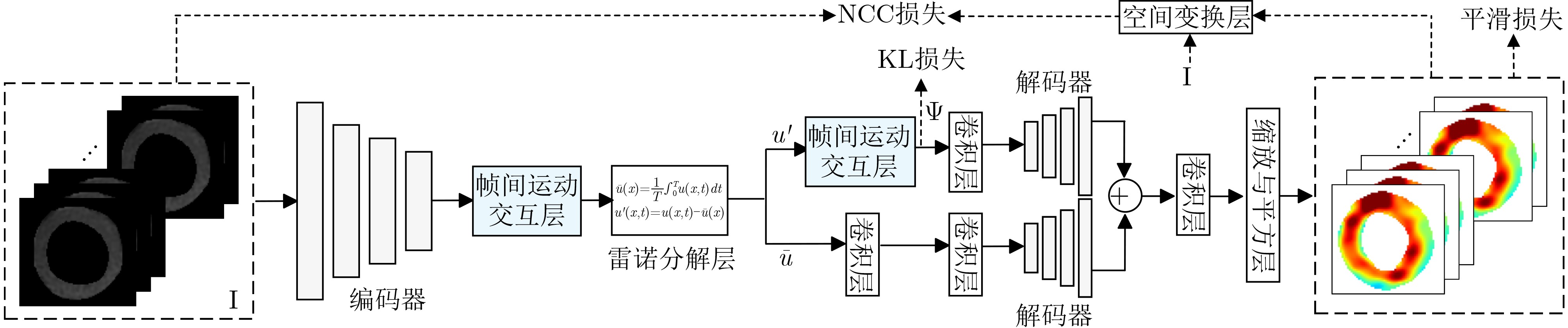
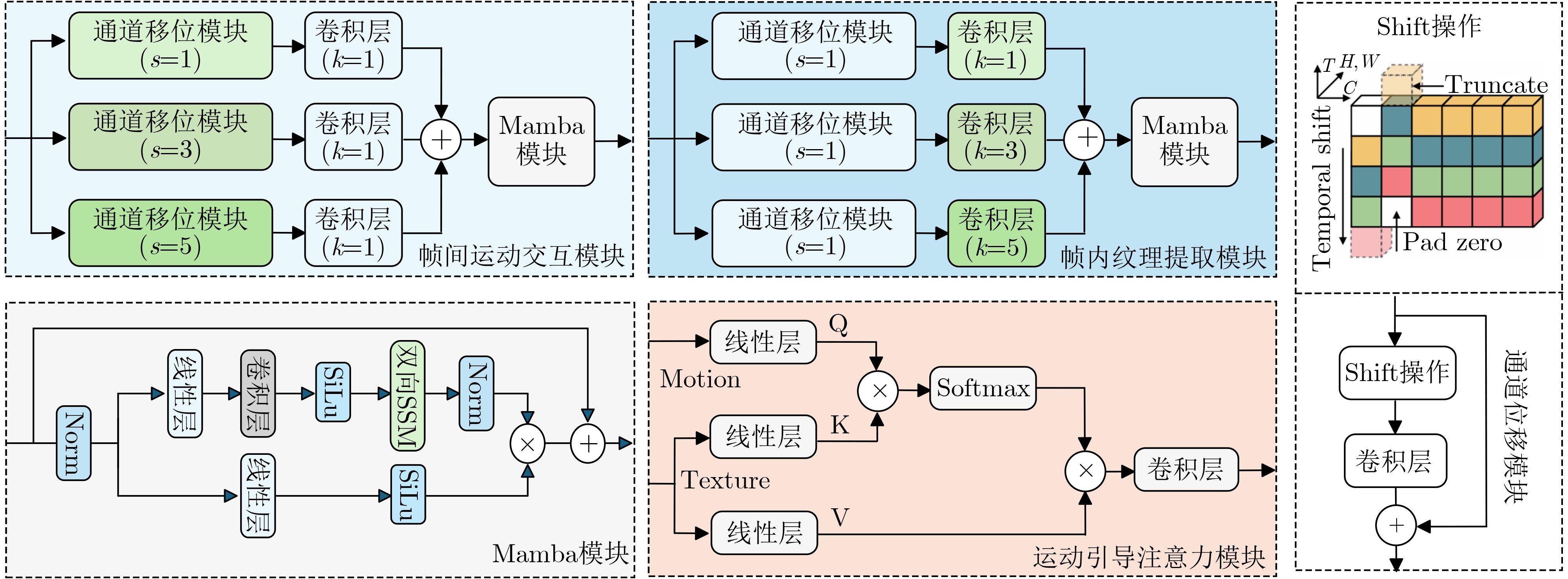
摘要: 针对现有基于心脏电影磁共振成像的瘢痕心肌分型方法中运动表征过于离散化且多模态融合机制难以挖掘深层病理关联的问题,该文提出一种基于运动引导纹理学习的瘢痕心肌分型方法(MGTNet)。该方法旨在通过精细化的运动场先验主动引导纹理特征的提取,实现运动与纹理的深度协同。首先引入流体力学中的雷诺分解思想构建雷诺分解运动网络,将心肌运动场解耦为规律性的平均运动与异常性的脉动运动以捕捉微小的运动异常。其次设计运动引导注意力模块,以提炼后的运动特征作为查询向量,通过交叉注意力机制定向增强纹理特征对病灶区域的感知能力。最后结合帧间运动交互与帧内纹理提取模块,实现端到端的精确分型。在CMRD与公开的ACDC数据集上的实验结果表明,该方法在各项关键指标上均优于现有的单模态及多模态融合方法。具体而言,MGTNet在两个数据集上的准确率分别达到95.6%和97.3%,AUC值均超过0.960。相比于基线网络运动与纹理网络(MTNet),该方法在准确率和F1分数上最高分别提升了1.4%和2.3%。结果证实MGTNet通过利用运动先验信息引导纹理特征提取的策略,有效提升了瘢痕心肌的无创诊断精度。Abstract:
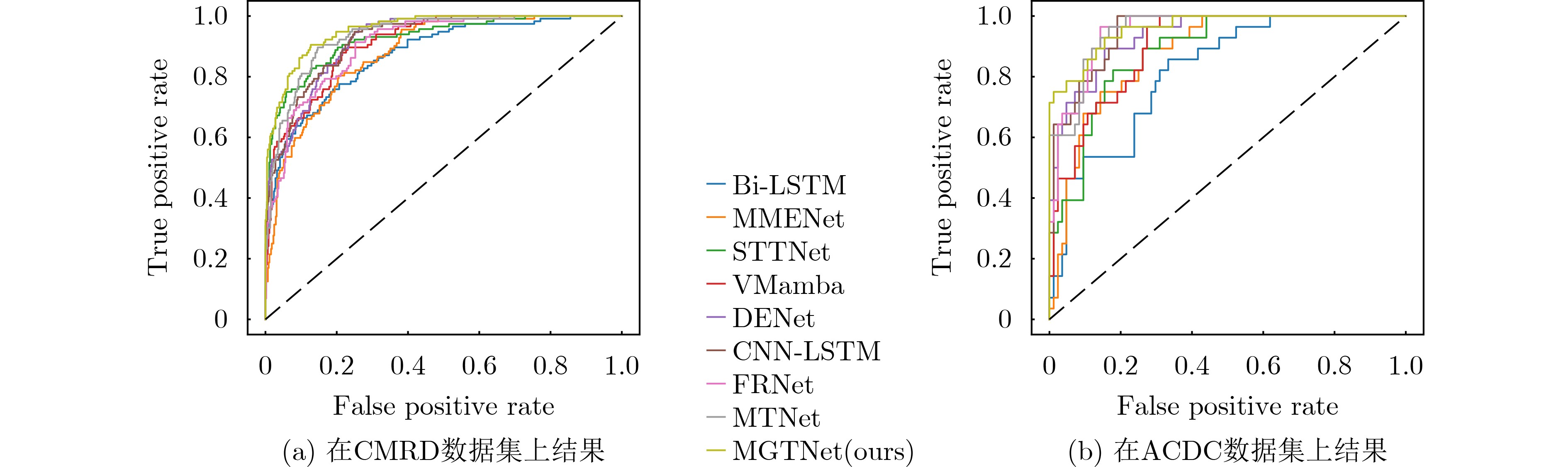
Objective Scarred myocardium is a key imaging marker of multiple cardiovascular diseases, and accurate phenotyping is clinically valuable for treatment planning and prognosis. Cine-MRI noninvasively provides both cardiac motion and anatomical texture information. However, existing classification methods have two major limitations. First, motion representation is often overly discretized and insensitive to subtle abnormalities. Second, multimodal fusion commonly relies on simple feature concatenation, which limits the ability to capture the deep pathological association between motion impairment and texture alteration. A motion-guided texture learning framework is therefore developed to improve the accuracy of non-invasive scarred myocardium classification. Methods A Motion-Guided Texture fusion Network (MGTNet) is proposed to enable deep interaction between motion and texture features. First, inspired by Reynolds decomposition in fluid dynamics, a Reynolds Decomposition Motion Network (RDMNet) is designed within a diffeomorphic registration framework to decompose the myocardial motion field into a regular mean motion component and an abnormal pulsatile motion component. This decomposition provides a refined motion representation that is sensitive to subtle abnormalities. Second, a Motion-Guided Cross-Attention (MGCA) module is designed, in which refined motion features serve as query features to dynamically modulate texture features and enhance the perception of suspected lesion regions. In addition, an inter-frame motion interaction module is used to capture temporal dependencies across cardiac frames, while an intra-frame texture extraction module learns multi-scale spatial texture patterns within each frame. Through a serial pipeline of motion field estimation, motion-guided texture enhancement, and feature fusion, end-to-end scarred myocardium classification is achieved. Results and Discussions Experiments on the CMRD and ACDC datasets show that MGTNet consistently outperforms existing single-modality and multimodal methods ( Tables 1 and2 ). It achieves accuracies of 95.6% on CMRD and 97.3% on ACDC, with AUC values of 96.6% and 96.0%, respectively. Compared with the baseline MTNet, MGTNet improves accuracy by up to 1.4 percentage points and the F1-score by up to 2.3 percentage points. Further comparisons with different motion field estimation methods (Tables 3 and4 ) show that RDMNet provides more discriminative motion priors and achieves the best overall performance on both datasets. These results indicate that fine-grained motion modeling and motion-guided texture enhancement effectively capture complementary pathological information from Cine-MRI. The ROC curves of the nine methods on both datasets further support the superior classification performance of the proposed method.Conclusions A scarred myocardium classification method based on motion-guided texture learning is presented. Reynolds decomposition is incorporated into motion field estimation to separate regular mean motion from abnormal pulsatile motion, and cross-attention is used to guide texture extraction with motion priors. This strategy addresses the limitations of discrete motion representation and shallow multimodal fusion. The results confirm that deep motion-texture interaction improves the accuracy and robustness of non-invasive scarred myocardium classification and provides an effective approach for Cine-MRI-based myocardial phenotyping. -
表 1 在CMRD数据集上对比9种方法的分类性能(%)
方法 准确率 精确率 敏感度 特异性 F1分数 AUC值 Bi-LSTM 85.5±1.8 56.5±3.0 54.4±3.8 90.4±1.1 53.0±3.2 88.5±1.7 MMENet 86.2±1.7 60.3±3.1 57.5±3.7 90.1±0.7 57.2±2.9 90.6±1.5 STTNet 91.0±1.0 75.2±2.3 71.0±3.0 92.9±0.3 71.3±2.0 93.7±1.0 VMamba 89.6±0.8 70.8±2.8 61.9±3.5 92.4±0.6 62.3±2.7 92.6±1.0 DENet 90.8±1.0 74.6±2.8 68.3±3.0 93.3±0.7 68.4±2.8 93.1±1.3 CNN-LSTM 91.9±0.8 78.5±2.5 74.7±2.5 94.2±0.5 74.6±2.3 93.8±1.1 FRNet 92.1±0.6 82.3±2.0 81.1±2.0 92.0±0.4 80.7±2.0 92.4±0.9 MTNet 94.2±0.4 86.1±1.7 87.5±1.8 95.9±0.3 86.8±1.8 95.1±0.8 MGTNet(本文) 95.6±0.4 88.7±1.5 86.9±1.7 96.3±0.3 89.1±1.7 96.6±0.7  下载: 导出CSV
表 2 在ACDC数据集上对比九种方法的分类性能(%)
方法 准确率 精确率 敏感度 特异性 F1分数 AUC值 Bi-LSTM 80.4±2.7 60.0±4.1 61.4±4.1 86.8±2.5 60.8±3.7 82.3±2.4 MMENet 78.6±2.7 64.4±3.8 60.7±4.6 85.5±2.7 59.6±3.5 81.8±2.6 STTNet 85.7±2.3 72.1±3.5 71.4±3.8 90.5±1.8 70.3±3.4 89.4±1.6 VMamba 91.1±1.8 82.3±3.2 82.0±2.6 94.1±1.3 81.1±3.4 89.8±1.7 DENet 92.5±1.5 84.6±2.8 85.0±2.4 95.2±1.1 84.0±2.9 91.2±1.4 CNN-LSTM 93.8±1.3 88.0±2.5 88.5±2.1 96.3±0.9 87.8±2.4 93.0±1.2 FRNet 93.4±1.0 92.0±1.8 91.2±1.7 96.3±1.0 90.1±2.0 93.5±1.0 MTNet 96.9±1.7 94.4±3.0 94.1±2.3 98.0±1.1 93.7±3.6 95.6±1.1 MGTNet(本文) 97.3±1.4 95.2±2.1 95.0±1.7 98.0±1.0 94.5±3.4 96.0±1.0
下载: 导出CSV
表 3 以MGTNet为网络主干,对比不同的运动场提取方法在CMRD数据集上的性能(%)
主干 运动 准确率 精确率 敏感度 特异性 F1分数 AUC值 MGTNet BOF 88.5±1.5 79.6±3.0 74.4±4.0 84.9±1.8 85.0±3.2 89.1±1.8 MEB 89.0±1.5 80.0±3.0 75.0±4.0 85.5±1.8 85.5±3.2 89.5±1.8 MQS 85.2±1.8 75.0±3.5 70.0±4.5 83.0±2.0 80.0±3.5 86.0±2.0 Voxelmorph 94.0±0.8 86.0±2.0 84.0±2.0 95.0±0.6 87.0±1.8 95.0±0.9 Deeptag 95.0±0.5 88.0±1.5 86.0±1.7 96.4±0.3 88.8±1.6 96.2±0.7 TransMorph 94.3±0.7 86.5±1.8 84.5±1.8 95.3±0.5 87.5±1.7 95.3±0.8 CorrMLP 94.7±0.6 87.2±1.6 85.5±1.6 95.8±0.4 88.2±1.6 95.8±0.7 RDMNet(本文) 95.6±0.4 88.7±1.5 86.9±1.7 96.3±0.3 89.1±1.7 96.6±0.7
下载: 导出CSV
表 4 以MGTNet为网络主干,对比不同的运动场提取方法在ACDC数据集上的性能(%)
主干 运动 准确率 精确率 敏感度 特异性 F1分数 AUC值 MGTNet BOF 89.5±1.5 77.6±3.0 80.4±3.2 89.1±1.6 85.0±3.2 90.5±1.6 MEB 90.0±1.5 78.5±3.0 81.0±3.2 89.5±1.6 85.5±3.2 91.0±1.6 MQS 86.0±1.8 75.0±3.5 77.0±3.5 87.0±1.8 82.0±3.5 88.0±1.8 Voxelmorph 92.0±1.3 85.0±2.8 87.0±2.5 93.0±1.3 88.0±3.0 92.5±1.3 Deeptag 95.5±1.0 93.0±2.2 92.0±2.0 96.5±0.9 91.5±2.5 94.5±1.0 TransMorph 93.5±1.2 88.0±2.5 90.0±2.3 94.5±1.1 90.0±2.8 93.5±1.2 CorrMLP 96.5±0.6 93.5±2.0 94.5±1.5 97.5±0.5 93.5±2.0 95.8±0.6 RDMNet(本文) 97.3±1.6 95.2±2.8 95.0±2.1 98.0±1.0 94.5±3.4 96.0±1.0
下载: 导出CSV
-
[1] ZHAO Xiaoying, ZHAO Xinxiang, JIN Fuwei, et al. Prognostic value of cardiac-MRI scar heterogeneity combined with left ventricular strain in patients with myocardial infarction[J]. Journal of Magnetic Resonance Imaging, 2023, 58(2): 466–476. doi: 10.1002/jmri.28478. [2] SERMESANT M, DELINGETTE H, COCHET H, et al. Applications of artificial intelligence in cardiovascular imaging[J]. Nature Reviews Cardiology, 2021, 18(8): 600–609. doi: 10.1038/s41569-021-00527-2. [3] SCHUETT K, MARX N, and LEHRKE M. The cardio-kidney patient: Epidemiology, clinical characteristics and therapy[J]. Circulation Research, 2023, 132(8): 902–914. doi: 10.1161/CIRCRESAHA.122.321748. [4] CAPTUR G, MANISTY C, and MOON J C. Cardiac MRI evaluation of myocardial disease[J]. Heart, 2016, 102(18): 1429–1435. doi: 10.1136/heartjnl-2015-309077. [5] BERNARD O, LALANDE A, ZOTTI C, et al. Deep learning techniques for automatic MRI cardiac multi-structures segmentation and diagnosis: Is the problem solved?[J]. IEEE Transactions on Medical Imaging, 2018, 37(11): 2514–2525. doi: 10.1109/TMI.2018.2837502. [6] LARROZA A, LÓPEZ-LEREU M P, MONMENEU J V, et al. Texture analysis of cardiac cine magnetic resonance imaging to detect nonviable segments in patients with chronic myocardial infarction[J]. Medical Physics, 2018, 45(4): 1471–1480. doi: 10.1002/mp.12783. [7] XIE Saining, GIRSHICK R, DOLLÁR P, et al. Aggregated residual transformations for deep neural networks[C]. The 2017 IEEE Conference on Computer Vision and Pattern Recognition (CVPR), Honolulu, USA, 2017: 5987–5995. doi: 10.1109/CVPR.2017.634. [8] 张坤. 基于残差网络的胎儿先天性心脏病检测方法的研究与实现[D]. [硕士论文], 济南大学, 2024. doi: 10.27166/d.cnki.gsdcc.2024.000200.ZHANG Kun. Research and implementation of fetal congenital heart disease detection method based on residual networks[D]. [Master dissertation], University of Ji’nan, 2024. doi: 10.27166/d.cnki.gsdcc.2024.000200. [9] 喻坤. 基于深度学习的心肌病变影像智能分析方法研究[D]. [硕士论文], 北京邮电大学, 2022. doi: 10.26969/d.cnki.gbydu.2022.001496.YU Kun. Intelligent analysis of myocardial infarction image based on deep learning[D]. [Master dissertation], Beijing University of Posts and Telecommunications, 2022. doi: 10.26969/d.cnki.gbydu.2022.001496. [10] LIU Ze, LIN Yutong, CAO Yue, et al. Swin transformer: Hierarchical vision transformer using shifted windows[C]. The 2021 IEEE/CVF International Conference on Computer Vision (ICCV), Montreal, Canada, 2021: 9992–10002. doi: 10.1109/ICCV48922.2021.00986. [11] ORTIZ-GONZALEZ A, KOBLER E, SIMON S, et al. Optical flow-guided cine MRI segmentation with learned corrections[J]. IEEE Transactions on Medical Imaging, 2024, 43(3): 940–953. doi: 10.1109/tmi.2023.3325766. [12] DORAZIL J, FLEURY B H, and HLAWATSCH F. Bayesian methods for optical flow estimation using a variational approximation, with applications to ultrasound[C]. ICASSP 2023–2023 IEEE International Conference on Acoustics, Speech and Signal Processing (ICASSP), Rhodes Greece, 2023: 1–5. doi: 10.1109/ICASSP49357.2023.10095694. [13] DILEEP P, RAO K N, BODAPATI P, et al. An automatic heart disease prediction using cluster-based bi-directional LSTM (C-BiLSTM) algorithm[J]. Neural Computing and Applications, 2023, 35(10): 7253–7266. doi: 10.1007/s00521-022-07064-0. [14] BACCOUCH W, HADIDI T, BENAMEUR N, et al. Convolution neural network for objective myocardial viability assessment based on regional wall thickness quantification from cine-MR images[J]. IEEE Access, 2024, 12: 112381–112396. doi: 10.1109/access.2024.3442080. [15] AHMADI N, TSANG M Y, GU Angnan, et al. Transformer-based spatio-temporal analysis for classification of aortic stenosis severity from echocardiography cine series[J]. IEEE Transactions on Medical Imaging, 2024, 43(1): 366–376. doi: 10.1109/tmi.2023.3305384. [16] RAJIAH P S, KALISZ K, BRONCANO J, et al. Myocardial strain evaluation with cardiovascular MRI: Physics, principles, and clinical applications[J]. RadioGraphics, 2022, 42(4): 968–990. doi: 10.1148/rg.210174. [17] CHEN Xiang, XIA Yan, DALL’ARMELLINA E, et al. Joint shape/texture representation learning for cardiovascular disease diagnosis from magnetic resonance imaging[J]. European Heart Journal-Imaging Methods and Practice, 2024, 2(1): qyae042. doi: 10.1093/ehjimp/qyae042. [18] REYNOLDS O. XXIX. An experimental investigation of the circumstances which determine whether the motion of water shall be direct or sinuous, and of the law of resistance in parallel channels[J]. Philosophical Transactions of the Royal Society of London, 1883(174): 935–982. doi: 10.1098/rstl.1883.0029. [19] LAUNDER B E. First steps in modelling turbulence and its origins: A commentary on Reynolds (1895) ‘on the dynamical theory of incompressible viscous fluids and the determination of the criterion’[J]. Philosophical Transactions of the Royal Society A: Mathematical, Physical and Engineering Sciences, 2015, 373(2039): 20140231. doi: 10.1098/rsta.2014.0231. [20] SUN Bohua. Revisiting the Reynolds-averaged Navier–Stokes equations[J]. Open Physics, 2021, 19(1): 853–862. doi: 10.1515/phys-2021-0102. [21] DEISSLER R G. Turbulent fluid motion IV-averages, Reynolds decomposition, and the closure problem[R]. NAS 1.15: 105822, 1992. [22] CAO Yao, MILLER M I, WINSLOW R L, et al. Large deformation diffeomorphic metric mapping of vector fields[J]. IEEE Transactions on Medical Imaging, 2005, 24(9): 1216–1230. doi: 10.1109/TMI.2005.853923. [23] CHEN C F R, FAN Quanfu, and PANDA R. CrossViT: Cross-attention multi-scale vision transformer for image classification[C]. The 2021 IEEE/CVF International Conference on Computer Vision (ICCV), Montreal, Canada, 2021: 347–356. doi: 10.1109/ICCV48922.2021.00041. [24] CHEN Junyu, FREY E C, HE Yufan, et al. TransMorph: Transformer for unsupervised medical image registration[J]. Medical Image Analysis, 2022, 82: 102615. doi: 10.1016/j.media.2022.102615. [25] BALAKRISHNAN G, ZHAO A, SABUNCU M R, et al. VoxelMorph: A learning framework for deformable medical image registration[J]. IEEE Transactions on Medical Imaging, 2019, 38(8): 1788–1800. doi: 10.1109/TMI.2019.2897538. [26] ZHANG Zhuming, HU Yongtao, YU Guoxing, et al. DeepTag: A general framework for fiducial marker design and detection[J]. IEEE Transactions on Pattern Analysis and Machine Intelligence, 2022, 45(3): 2931–2944. doi: 10.1109/tpami.2022.3174603. [27] LIN Ji, GAN Chuang, and HAN Song. TSM: Temporal shift module for efficient video understanding[C]. The 2019 IEEE/CVF International Conference on Computer Vision (ICCV), Seoul, Korea, 2019: 7082–7092. doi: 10.1109/ICCV.2019.00718. [28] LIU Yue, TIAN Yunjie, ZHAO Yuzhong, et al. VMamba: Visual state space model[C]. The 38th International Conference on Neural Information Processing Systems, Vancouver, Canada, 2024: 3273. [29] WANG Yaming, YANG Daiguo, PU Cailing, et al. Combined myocardial motion and texture characterisation methods for the phenotyping of scarred myocardium[J]. Expert Systems with Applications, 2026, 296: 128936. doi: 10.1016/j.eswa.2025.128936. [30] PAN Jiazhen, HUANG Wenqi, RUECKERT D, et al. Motion-compensated MR CINE reconstruction with reconstruction-driven motion estimation[J]. IEEE Transactions on Medical Imaging, 2024, 43(7): 2420–2433. doi: 10.1109/TMI.2024.3364504. [31] 邱麒添. 基于CNN-LSTM混合模型的心脏病风险分类预测研究[J]. 电脑知识与技术, 2025, 21(30): 21–23. doi: 10.14004/j.cnki.ckt.2025.1511.QIU Qitian. Research on risk classification and prediction of heart disease based on CNN-LSTM hybrid model[J]. Computer Knowledge and Technology, 2025, 21(30): 21–23. doi: 10.14004/j.cnki.ckt.2025.1511. [32] TIAN Qiuhong, MIAO Weilun, ZHANG Lizao, et al. MCANet: A lightweight action recognition network with multidimensional convolution and attention[J]. International Journal of Machine Learning and Cybernetics, 2025, 16(5): 3345–3358. doi: 10.1007/s13042-024-02454-3. [33] MENG Mingyuan, FENG Dagan, BI Lei, et al. Correlation-aware coarse-to-fine MLPs for deformable medical image registration[C]. The 2024 IEEE/CVF Conference on Computer Vision and Pattern Recognition (CVPR), Seattle, USA, 2024: 9645–9654. doi: 10.1109/CVPR52733.2024.00921. [34] 陈晓禾, 张甲昂, 李玲芝, 等. 面向病理图像弱监督分类的层级融合多实例学习方法[J]. 电子与信息学报, 2026, 48(3): 1116–1127. doi: 10.11999/JEIT250726.CHEN Xiaohe, ZHANG Jia’ang, LI Lingzhi, et al. Hierarchical fusion multi-instance learning for weakly supervised pathological image classification[J]. Journal of Electronics & Information Technology, 2026, 48(3): 1116–1127. doi: 10.11999/JEIT250726. [35] 崔学英, 王宇航, 刘斌, 等. Wave-MambaCT: 基于小波Mamba的低剂量CT伪影抑制方法[J]. 电子与信息学报, 2026, 48(3): 982–993. doi: 10.11999/JEIT250489.CUI Xueying, WANG Yuhang, LIU Bin, et al. Wave-MambaCT: Low-dose CT artifact suppression method based on wavelet Mamba[J]. Journal of Electronics & Information Technology, 2026, 48(3): 982–993. doi: 10.11999/JEIT250489. [36] 张印辉, 刘凯, 何自芬, 等. 动态小波多向感知与几何轴解引导三维CT骨折图像分割[J]. 电子与信息学报, 2026, 48(3): 1005–1016. doi: 10.11999/JEIT250732.ZHANG Yinhui, LIU Kai, HE Zifen, et al. Dynamic wavelet multi-directional perception and geometry axis-solution guided 3D CT fracture image segmentation[J]. Journal of Electronics & Information Technology, 2026, 48(3): 1005–1016. doi: 10.11999/JEIT250732. -

 下载:
下载:


图(6) / 表(4)
计量
- 文章访问数: 237
- HTML全文浏览量: 55
- PDF下载量: 13
- 被引次数: 0
