




MGM-3DUNet: A Multi-scale Edge Semantic-guided GraphConvolutional Sequence Method for Brain Tumor Segmentation
-
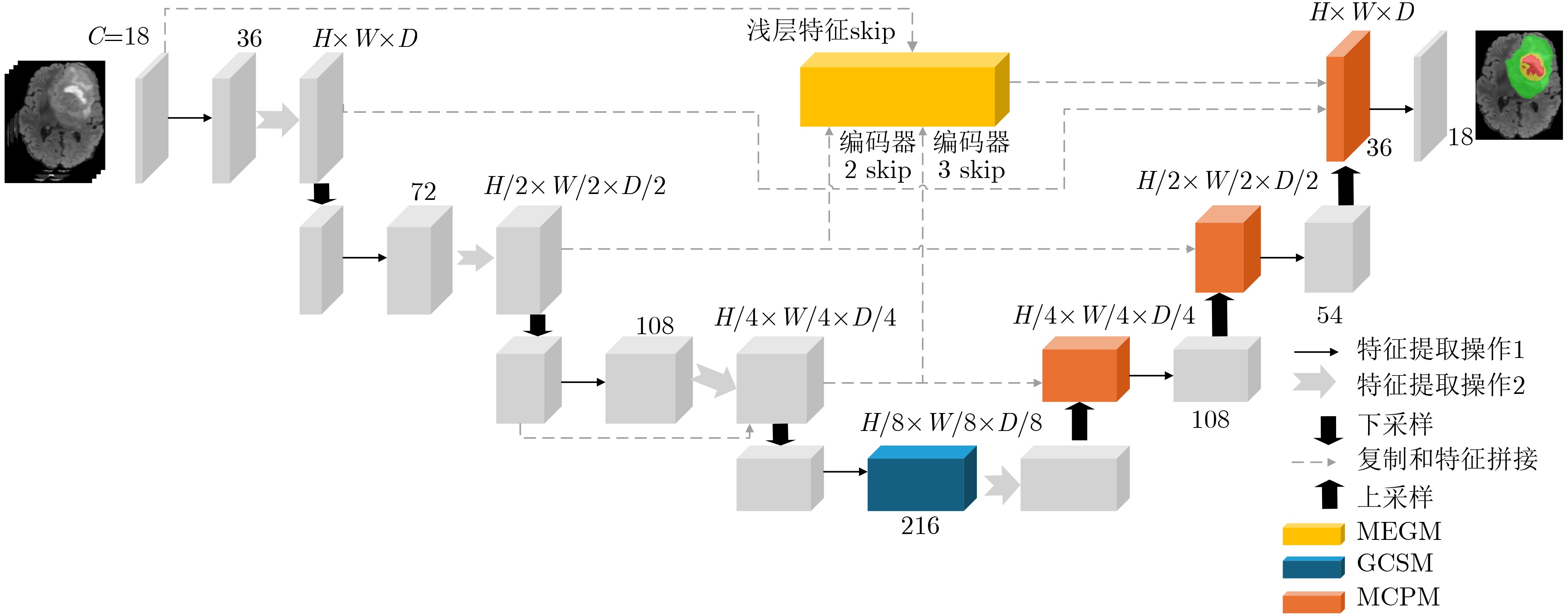
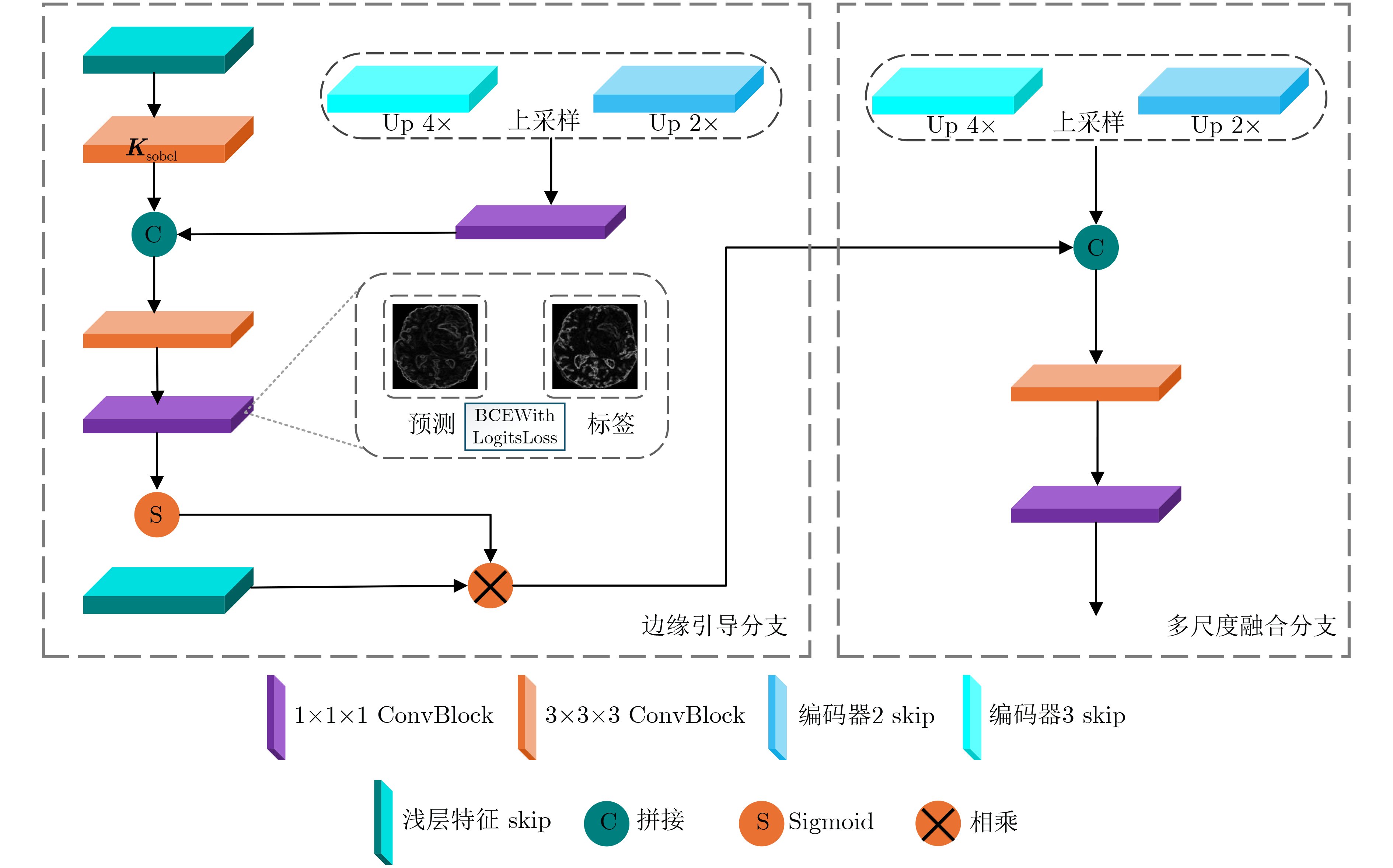
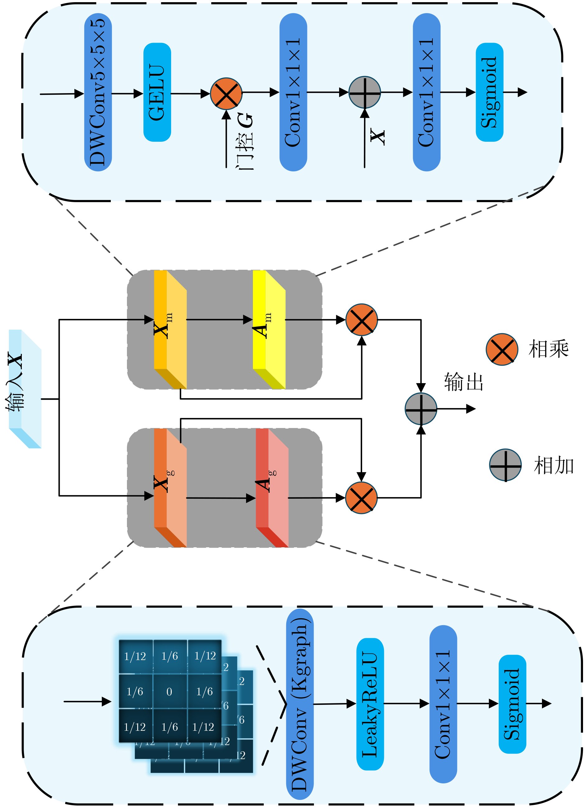
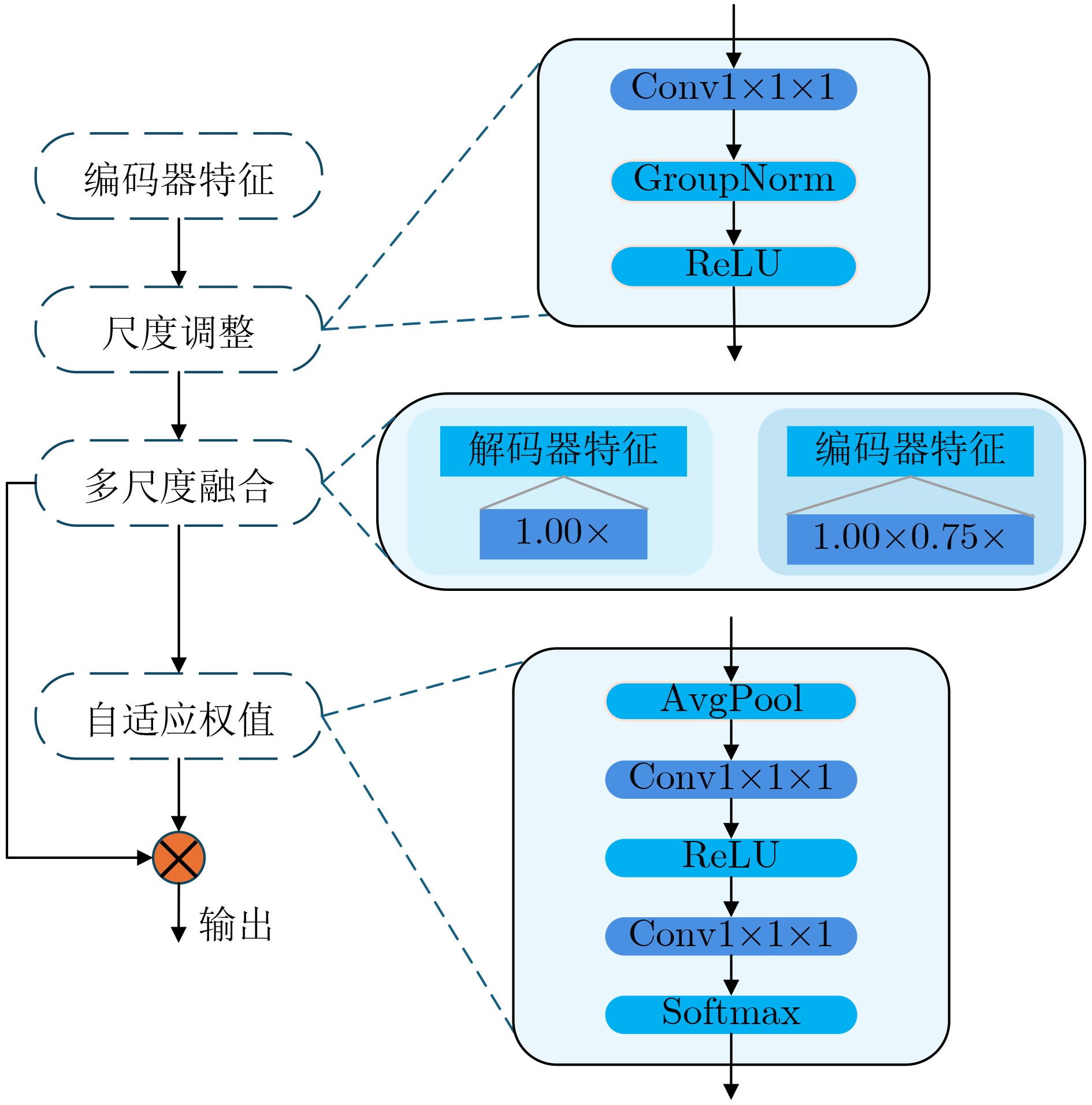
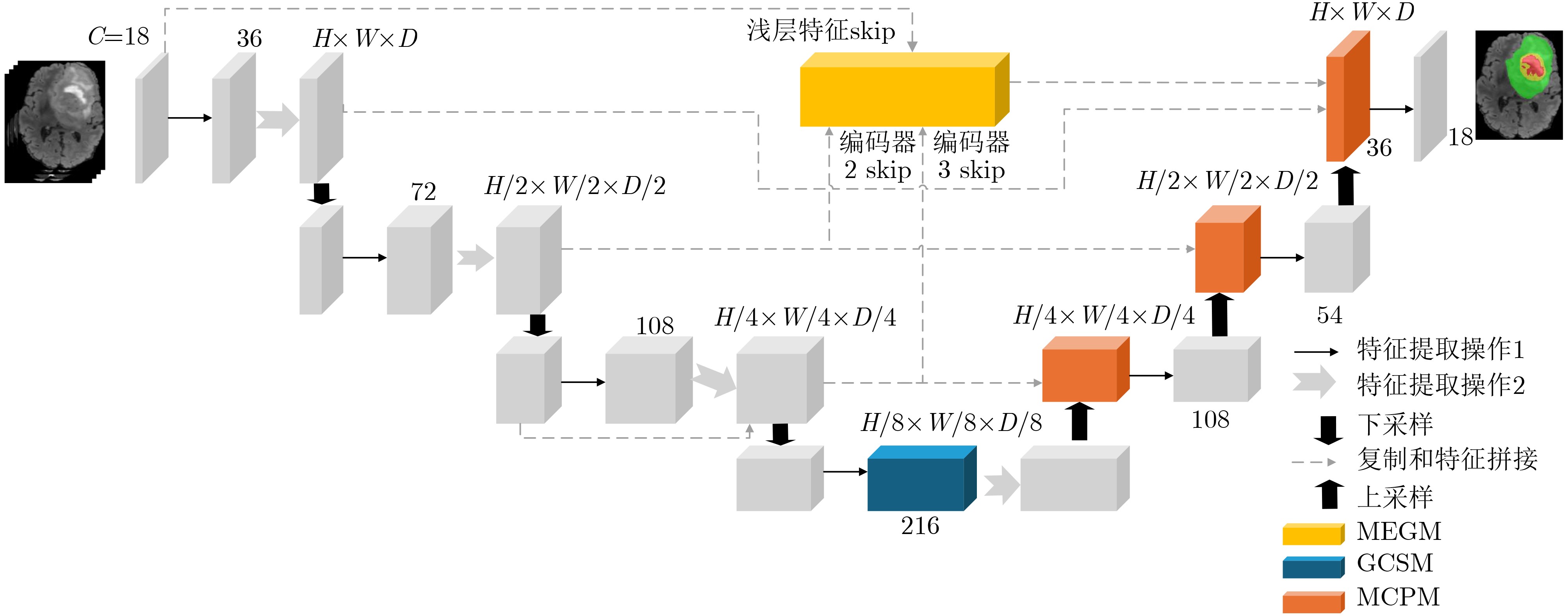
摘要: 针对脑肿瘤尺度差异大、边界模糊、小病灶易漏检以及现有分割模型难以兼顾分割性能和参数规模等问题,该文以三维U型卷积神经网络(3DUNet)为基线,提出多尺度边缘语义引导的图卷积序列脑肿瘤分割方法—MGM-3DUNet。通过多尺度边缘语义引导模块(MEGM)将可学习的边缘检测与多尺度语义特征融合,精准捕捉肿瘤边界细节,生成边缘预测图提供辅助监督;通过图卷积序列模块(GCSM)融合图卷积的局部拓扑聚合能力与高效长程建模优势,缓解现有长序列特征建模方法在特征编码过程中局部细粒度细节易衰减、空间精细结构保留不足的问题;多尺度上下文感知模块(MCPM)对不同尺度特征动态加权融合,使解码器自适应匹配水肿区与核心区的特征需求,缓解尺度失衡导致的小病灶漏检。最后,在开源数据集BraTS2020, BraTS2021上进行了实验验证。结果表明,模型以2.3 M的轻量级参数在整体肿瘤(WT)、肿瘤核心(TC)和增强肿瘤(ET)区域分别达到了91.2%, 90.4%和89.2%的Dice系数,整体性能优于现有主流模型,在保持低计算成本的同时,实现了关键区域的精准分割。Abstract:
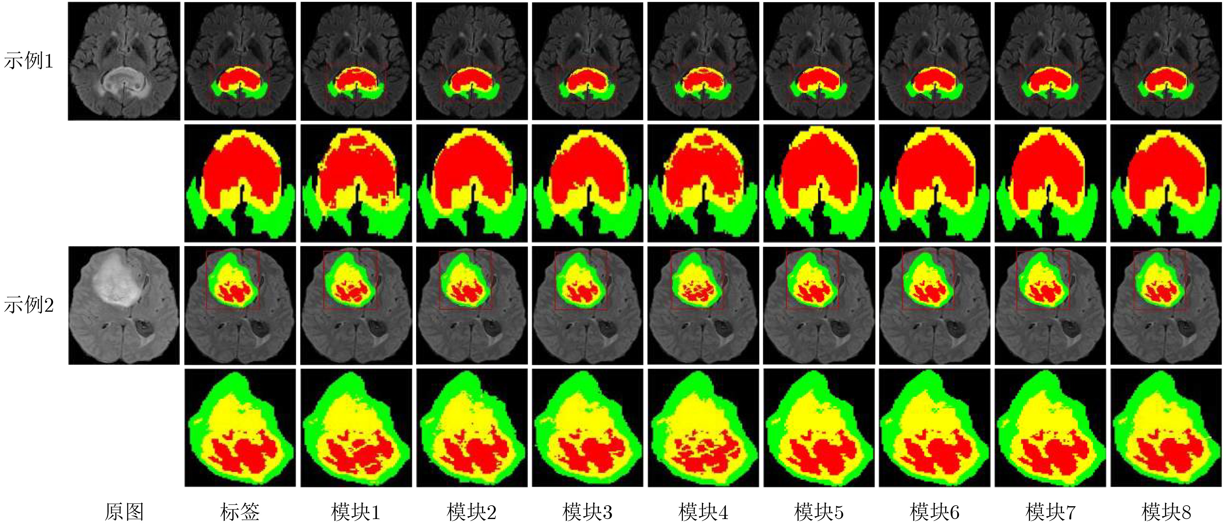
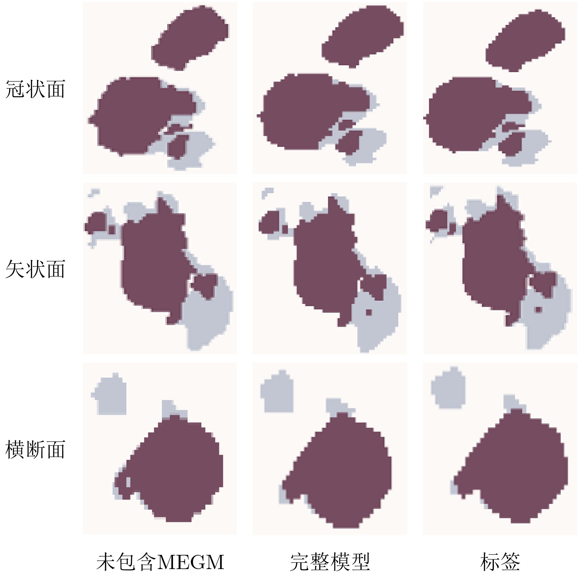
Objective Feature fusion in U-Net and its 3D variants mainly relies on simple single-scale concatenation, which limits the use of encoder features and weakens fine-grained segmentation of Tumor Core (TC) and Enhancing Tumor (ET) regions. Recent methods such as VM-UNet improve sequence modeling efficiency, but they mainly focus on global information modeling. Local detail preservation and edge enhancement remain insufficient. Therefore, current methods still have limitations in segmentation accuracy and clinical utility. To address these problems, this paper proposes MGM-3DUNet for brain tumor segmentation. Methods The Multi-scale Edge semantic Guidance Module (MEGM) is designed to improve tumor boundary segmentation through learnable edge detection. The Graph Convolutional Sequence Module (GCSM) combines the local aggregation ability of graph convolution with efficient long-range modeling based on a Mamba-like structure. This design improves semantic consistency while preserving small tumor structures with fewer parameters. The Multi-scale Context Perception Module (MCPM) is introduced to strengthen feature complementarity across different tumor scales through dual-scale fusion. Results and Discussions Experiments show that the proposed method achieves better average Dice similarity coefficient (Dice) and 95th percentile Hausdorff Distance (HD95) than the comparison methods. With only 2.3 M parameters, MGM-3DUNet achieves Dice values of 91.2%, 90.4%, and 89.2% for Whole Tumor (WT), TC, and ET, respectively. The visualization results ( Fig. 9 ,Fig. 10 ) further show that MEGM improves boundary localization. Overall, the proposed method shows improved sensitivity to edge details and contextual correlations while maintaining a low parameter count.Conclusions This method improves tumor boundary prediction by introducing shallow-layer edge enhancement to emphasize tumor contours. Local and global semantic information is fused in the bottleneck layer, and multi-scale contextual features are integrated during decoding. The proposed design achieves accurate segmentation with low computational cost and is suitable for deployment on resource-constrained platforms. -
Key words:
- 3DUNet /
- Lightweight /
- Edge semantic guidance /
- Graph convolution /
- Brain tumor segmentation
-
表 1 实验设备配置表
参数 配置 CPU Intel(R)Core(TM)i7-14700HX(2.1 GHz) GPU NVIDIA GeForce RTX 4060(8 GB) Windows 11 CUDA 11.8 PyTorch 2.5 Python 3.9  下载: 导出CSV
表 2 数据集划分及样本量分布
数据集名称 数据子集 样本数量 用途 BraTS2021 训练集 875 模型训练 验证集 250 性能验证 测试集 125 性能测试 BraTS2020 测试集 50 泛化验证
下载: 导出CSV
表 3 在BraTS2021数据集上使用基线模型3DUNet不同$ \alpha $时的结果对比
$ \alpha $ Dice(%) Sensitivity(%) HD95(mm) WT TC ET 均值 WT TC ET 均值 WT TC ET 均值 0.1 83.3 75.4 74.2 77.6 88.3 78.2 73.2 79.9 18.6 12.1 10.1 13.6 0.2 82.1 75.6 75.3 77.7 87.5 78.1 75.3 80.3 17.5 11.5 10.3 13.1 0.3 84.8 76.3 75.6 78.9 89.7 80.6 77.0 82.4 16.9 10.9 9.9 12.6 0.4 81.2 76.4 72.1 76.6 86.7 79.9 73.1 79.9 18.3 10.9 10.3 13.2 0.5 80.5 75.8 73.2 76.5 86.3 78.2 72.8 79.1 18.2 12.3 10.8 13.8 0.6 82.5 74.7 74.4 77.2 87.9 77.8 75.9 80.5 17.8 12.5 10.5 13.6 0.7 83.6 74.2 74.1 77.3 88.5 77.2 75.2 80.3 17.1 12.4 10.4 13.3 0.8 80.2 75.5 73.9 76.5 85.4 77.3 73.0 78.6 18.8 11.8 10.7 13.8 0.9 79.8 73.1 73.6 75.5 84.9 75.4 74.6 78.3 19.0 12.7 11.0 14.2 注:表中粗体表示最优值。
下载: 导出CSV
表 4 交叉验证结果对比
交叉验证折数 Dice(%) Sensitivity(%) HD95(mm) WT TC ET 均值 WT TC ET 均值 WT TC ET 均值 1 93.1 90.4 90.2 91.2 96.2 96.1 89.9 94.1 2.8 2.1 3.0 2.6 2 92.1 88.9 88.6 89.9 95.5 92.4 88.3 92.1 3.2 2.7 3.3 3.1 3 93.8 93.3 89.3 92.1 96.7 95.6 89.0 93.8 3.0 2.9 2.9 2.9 4 91.2 90.2 89.4 90.3 93.8 92.9 89.1 91.9 3.1 2.9 3.7 3.2 5 92.5 89.8 88.2 90.2 95.3 92.2 88.3 91.9 2.9 2.3 3.4 2.9 均值 92.5 90.5 89.1 90.7 95.5 93.8 88.9 92.7 3.0 2.6 3.3 3.0 注:表中所有数值均为平均值。
下载: 导出CSV
表 5 消融实验结果对比
模块 Dice(%) Sensitivity(%) HD95(mm) WT TC ET 均值 WT TC ET 均值 WT TC ET 均值 3DUNet(1) 82.3 76.1 75.3 77.9 88.1 81.2 75.8 81.7 17.7 11.4 9.5 12.9 3DUNet+MEGM(2) 89.8 87.2 86.8 87.9 94.8 88.6 82.2 88.5 7.2 5.5 5.3 6.0 3DUNet+GCSM(3) 88.4 85.0 85.8 86.4 94.3 87.8 84.4 88.8 8.4 7.2 5.1 6.9 3DUNet+MCPM(4) 86.2 83.1 80.5 83.3 92.8 85.5 79.4 85.9 11.7 9.2 8.3 9.7 3DUNet+MEGM+GCSM(5) 90.2 88.8 88.2 89.1 95.3 90.2 89.8 91.8 6.4 4.5 4.2 5.0 3DUNet+MEGM+MCPM(6) 89.2 87.4 87.6 88.1 95.0 88.9 87.1 90.3 5.9 4.3 3.9 4.7 3DUNet+GCSM+MCPM(7) 88.9 88.2 86.2 87.8 94.2 88.2 88.6 90.3 6.2 5.1 4.5 5.3 MGM-3DUNet(8) 91.2 90.4 89.2 90.3 96.4 93.2 90.2 93.3 3.1 3.9 2.8 3.3 注:表中粗体表示最优值。
下载: 导出CSV
表 6 MGM-3DUNet与不同模型的对比结果
网络类型 模型 Dice(%) HD95(%) Param(M) WT TC ET 均值 WT TC ET 均值 经典网络 UNet++[22] 86.6 77.4 74.5 79.5 8.9 23.3 27.9 20.0 22.2 V-Net[6] 88.2 83.6 80.6 84.1 10.5 13.3 24.5 16.1 24.3 MGM-3DUNet 91.2 90.4 89.2 90.3 3.1 3.9 2.8 3.3 2.3 目前主流网络 TransBTS[9] 86.4 88.3 87.6 87.4 12.5 16.8 13.1 14.1 30.6 UNETR[23] 90.8 89.3 88.9 89.7 5.5 5.0 3.6 4.7 148.5 Swin UNETR[11] 90.8 89.5 89.0 89.8 5.4 4.8 3.7 4.6 35.5 VM-UNet[15] 90.2 85.6 80.2 85.3 5.4 7.8 5.3 6.2 18.7 FS Inv-ResU-Net[24] 90.5 86.5 82.8 86.6 5.5 4.5 2.4 4.1 26.3 MR-SC-UNet[25] 91.1 87.5 87.8 88.8 4.4 5.3 2.5 4.1 32.4 DC-Seg[26] 89.8 88.2 87.9 88.6 5.8 6.2 5.2 5.7 15.2 VSMU-Net[27] 90.4 89.3 88.6 89.4 6.1 4.8 5.0 5.3 25.1 S2CA-Net[28] 89.9 89.2 88.5 89.2 6.3 5.1 4.9 5.4 32.3 MGM-3DUNet 91.2 90.4 89.2 90.3 3.1 3.9 2.8 3.3 2.3 轻量级网络 LATUP-Net[29] 90.5 88.5 86.4 88.5 6.3 8.2 4.5 6.3 3.1 ADHDC-Net[30] 80.5 87.0 85.4 84.3 14.7 12.1 6.8 11.2 0.3 MGM-3DUNet 91.2 90.4 89.2 90.3 3.1 3.9 2.8 3.3 2.3 注:表中粗体表示最优值
下载: 导出CSV
表 7 MGM-3DUNet在BraTS2020数据集上的对比结果(%)
模型 Dice HD95 WT TC ET 均值 WT TC ET 均值 VM-UNet 87.2 85.9 82.4 85.2 6.2 8.3 6.4 7.0 LATUP-Net 87.5 88.1 85.3 87.0 5.5 9.2 5.3 6.7 TransBTS 82.5 83.6 81.8 82.6 15.5 20.3 6.8 14.2 DC-Seg 89.2 88.1 86.7 88.0 7.3 6.1 5.5 6.3 VSMU-Net 89.5 88.3 87.1 88.3 7.1 5.7 5.5 6.1 S2CA-Net 89.1 88.1 87.2 88.1 7.2 6.3 5.4 6.3 MGM-3DUNet 90.1 89.8 87.2 89.0 5.2 4.8 4.0 4.7 注:表中粗体表示最优值
下载: 导出CSV
-
[1] KRISHNAPRIYA S and KARUNA Y. A survey of deep learning for MRI brain tumor segmentation methods: Trends, challenges, and future directions[J]. Health and Technology, 2023, 13(2): 181–201. doi: 10.1007/s12553-023-00737-3. [2] 李锵, 阮方号, 关欣. 基于双路径特征融合的轻量级脑肿瘤分割网络[J]. 天津大学学报: 自然科学与工程技术版, 2024, 57(11): 1177–1186. doi: 10.11784/tdxbz202401015.LI Qiang, RUAN Fanghao, and GUAN Xin. Lightweight brain tumor segmentation network based on dual-path feature fusion[J]. Journal of Tianjin University: Science and Technology, 2024, 57(11): 1177–1186. doi: 10.11784/tdxbz202401015. [3] 王晨, 杜晨曦, 刘瑞军, 等. 基于RGB-D图像的语义分割方法综述[J]. 计算机辅助设计与图形学学报, 2025, 37(1): 100–119. doi: 10.3724/SP.J.1089.2024-00107.WANG Chen, DU Chenxi, LIU Ruijun, et al. A review of semantic segmentation methods based on RGB-D images[J]. Journal of Computer-Aided Design & Computer Graphics, 2025, 37(1): 100–119. doi: 10.3724/SP.J.1089.2024-00107. [4] RONNEBERGER O, FISCHER P, and BROX T. U-Net: Convolutional networks for biomedical image segmentation[C]. The 18th International Conference on Medical Image Computing and Computer-Assisted Intervention, Munich, Germany, 2015: 234–241. doi: 10.1007/978-3-319-24574-4_28. [5] ÇIÇEK Ö, ABDULKADIR A, LIENKAMP S S, et al. 3D U-Net: Learning dense volumetric segmentation from sparse annotation[C]. The 19th International Conference on Medical Image Computing and Computer-Assisted Intervention, Athens, Greece, 2016: 424–432. doi: 10.1007/978-3-319-46723-8_49. [6] MILLETARI F, NAVAB N, and AHMADI S A. V-Net: Fully convolutional neural networks for volumetric medical image segmentation[C]. The 2016 Fourth International Conference on 3D Vision, Stanford, USA, 2016: 565–571. doi: 10.1109/3DV.2016.79. [7] VASWANI A, SHAZEER N, PARMAR N, et al. Attention is all you need[C]. The 31st International Conference on Neural Information Processing Systems, Long Beach, USA, 2017: 5998–6008. [8] DOSOVITSKIY A, BEYER L, KOLESNIKOV A, et al. An image is worth 16×16 words: Transformers for image recognition at scale[C]. The 9th International Conference on Learning Representations, 2021. [9] WANG Wenxuan, CHEN Chen, DING Meng, et al. TransBTS: Multimodal brain tumor segmentation using Transformer[C]. The 24th International Conference on Medical Image Computing and Computer-Assisted Intervention, Strasbourg, France, 2021: 109–119. doi: 10.1007/978-3-030-87193-2_11. [10] LIU Ze, LIN Yutong, CAO Yue, et al. Swin transformer: Hierarchical vision transformer using shifted windows[C]. 2021 IEEE/CVF International Conference on Computer Vision (ICCV), Montreal, Canada, 2021: 9992–10002. doi: 10.1109/ICCV48922.2021.00986. [11] HATAMIZADEH A, NATH V, TANG Yucheng, et al. Swin UNETR: Swin transformers for semantic segmentation of brain tumors in MRI images[C]. The 7th International Workshop Brainlesion: Glioma, Multiple Sclerosis, Stroke and Traumatic Brain Injuries, 2022: 272–284. doi: 10.1007/978-3-031-08999-2_22. [12] HE Kelei, GAN Chen, LI Zhuoyuan, et al. Transformers in medical image analysis[J]. Intelligent Medicine, 2023, 3(1): 59–78. doi: 10.1016/j.imed.2022.07.002. [13] 陈雷, 杨吉斌, 曹铁勇, 等. 一种基于Transformer特征金字塔的自蒸馏目标分割方法[J]. 电子与信息学报, 2025, 47(2): 551–560. doi: 10.11999/JEIT240735.CHEN Lei, YANG Jibin, CAO Tieyong, et al. A self-distillation object segmentation method based on Transformer feature pyramid[J]. Journal of Electronics & Information Technology, 2025, 47(2): 551–560. doi: 10.11999/JEIT240735. [14] MA Jun, LI Feifei, and WANG Bo. U-Mamba: Enhancing long-range dependency for biomedical image segmentation[EB/OL]. https://arxiv.org/abs/2401.04722, 2024. [15] RUAN Jiacheng, LI Jincheng, XIANG Suncheng, et al. VM-UNet: Vision mamba UNet for medical image segmentation[J]. ACM Transactions on Multimedia Computing, Communications and Applications, 2025. doi: 10.1145/3767748. [16] 丁建睿, 张听, 刘家栋, 等. 融合邻域注意力和状态空间模型的医学视频分割算法[J]. 电子与信息学报, 2025, 47(5): 1582–1595. doi: 10.11999/JEIT240755.DING Jianrui, ZHANG Ting, LIU Jiadong, et al. A medical video segmentation algorithm integrating neighborhood attention and state space model[J]. Journal of Electronics & Information Technology, 2025, 47(5): 1582–1595. doi: 10.11999/JEIT240755. [17] 孙军梅, 葛青青, 李秀梅, 等. 一种具有边缘增强特点的医学图像分割网络[J]. 电子与信息学报, 2022, 44(5): 1643–1652. doi: 10.11999/JEIT210784.SUN Junmei, GE Qingqing, LI Xiumei, et al. A medical image segmentation network with boundary enhancement[J]. Journal of Electronics & Information Technology, 2022, 44(5): 1643–1652. doi: 10.11999/JEIT210784. [18] 周涛, 党培, 陆惠玲, 等. 跨模态跨尺度跨维度的PET/CT图像的Transformer分割模型[J]. 电子与信息学报, 2023, 45(10): 3529–3537. doi: 10.11999/JEIT221204.ZHOU Tao, DANG Pei, LU Huiling, et al. A Transformer segmentation model for PET/CT images with cross-modal, cross-scale and cross-dimensional[J]. Journal of Electronics & Information Technology, 2023, 45(10): 3529–3537. doi: 10.11999/JEIT221204. [19] SUDRE C H, LI Wenqi, VERCAUTEREN T, et al. Generalised dice overlap as a deep learning loss function for highly unbalanced segmentations[C]. The 3rd International Workshop Deep Learning in Medical Image Analysis and Multimodal Learning for Clinical Decision Support, Québec City, Canada, 2017: 240–248. doi: 10.1007/978-3-319-67558-9_28. [20] 马金林, 杨继鹏. 基于TT分解的轻量化肝肿瘤分割方法[J]. 电子与信息学报, 2026, 48(1): 335–345. doi: 10.11999/JEIT250293.MA Jinlin and YANG Jipeng. Tensor-train decomposition for lightweight liver tumor segmentation[J]. Journal of Electronics & Information Technology, 2026, 48(1): 335–345. doi: 10.11999/JEIT250293. [21] 周涛, 叶鑫宇, 刘凤珍, 等. 利用跨模态轻量级YOLOv5模型的PET/CT肺部肿瘤检测[J]. 电子与信息学报, 2024, 46(2): 624–632. doi: 10.11999/JEIT230052.ZHOU Tao, YE Xinyu, LIU Fengzhen, et al. CL-YOLOv5: PET/CT lung cancer detection with cross-modal lightweight YOLOv5 model[J]. Journal of Electronics & Information Technology, 2024, 46(2): 624–632. doi: 10.11999/JEIT230052. [22] ZHOU Zongwei, RAHMAN SIDDIQUEE M, TAJBAKHSH N, et al. UNet++: A nested U-Net architecture for medical image segmentation[C]. The 4th Deep Learning in Medical Image Analysis and Multimodal Learning for Clinical Decision Support, Granada, Spain, 2018: 3–11. doi: 10.1007/978-3-030-00889-5_1. [23] HATAMIZADEH A, TANG Yucheng, NATH V, et al. UNETR: Transformers for 3D medical image segmentation[C]. The 2022 IEEE/CVF Winter Conference on Applications of Computer Vision, Waikoloa, USA, 2022: 1748–1758. doi: 10.1109/WACV51458.2022.00181. [24] 阮东升, 施哲彬, 王嘉辉, 等. 结合跨模态特征激励与双分支交叉注意力融合的左心房疤痕分割方法[J]. 电子与信息学报, 2025, 47(5): 1596–1608. doi: 10.11999/JEIT240775.RUAN Dongsheng, SHI Zhebin, WANG Jiahui, et al. Left atrial scar segmentation method combining cross-modal feature excitation and dual branch cross attention fusion[J]. Journal of Electronics & Information Technology, 2025, 47(5): 1596–1608. doi: 10.11999/JEIT240775. [25] LIU Jiangjiang, HOU Qibin, LIU Zhiang, et al. PoolNet+: Exploring the potential of pooling for salient object detection[J]. IEEE Transactions on Pattern Analysis and Machine Intelligence, 2023, 45(1): 887–904. doi: 10.1109/TPAMI.2021.3140168. [26] LI Haitao, LI Ziyu, MAO Yiheng, et al. DC-Seg: Disentangled contrastive learning for brain tumor segmentation with missing modalities[C]. The 28th International Conference on Medical Image Computing and Computer Assisted Intervention, Daejeon, South Korea, 2026: 138–148. doi: 10.1007/978-3-032-04984-1_14. [27] ZHU Zhiqin, WANG Zhandong, QI Guanqiu, et al. Visually stabilized Mamba U-shaped network with strong inductive bias for 3-D brain tumor segmentation[J]. IEEE Transactions on Instrumentation and Measurement, 2025, 74: 2518511. doi: 10.1109/TIM.2025.3551581. [28] ZHOU Lifang, JIANG Yu, LI Weisheng, et al. Shape-scale co-awareness network for 3D brain tumor segmentation[J]. IEEE Transactions on Medical Imaging, 2024, 43(7): 2495–2508. doi: 10.1109/TMI.2024.3368531. [29] ALWADEE E J, SUN Xianfang, QIN Yipeng, et al. LATUP-Net: A lightweight 3D attention U-Net with parallel convolutions for brain tumor segmentation[J]. Computers in Biology and Medicine, 2025, 184: 109353. doi: 10.1016/j.compbiomed.2024.109353. [30] LIU Hengxin, HUO Guoqiang, LI Qiang, et al. Multiscale lightweight 3D segmentation algorithm with attention mechanism: Brain tumor image segmentation[J]. Expert Systems with Applications, 2023, 214: 119166. doi: 10.1016/j.eswa.2022.119166. -

 下载:
下载:


图(11) / 表(7)
计量
- 文章访问数: 513
- HTML全文浏览量: 134
- PDF下载量: 58
- 被引次数: 0
