

A Contour Detection Method Based on Interactive Perception Mechanism of Dual Visual Pathways
-
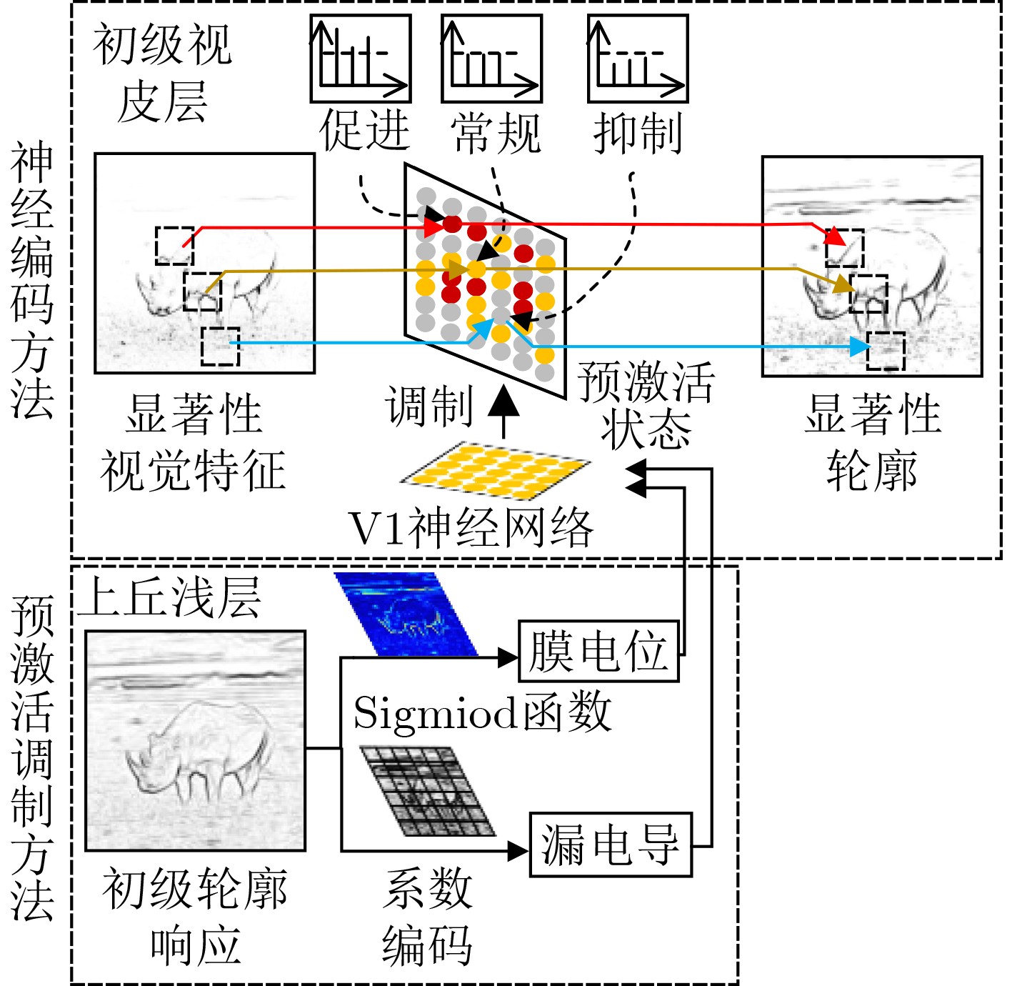
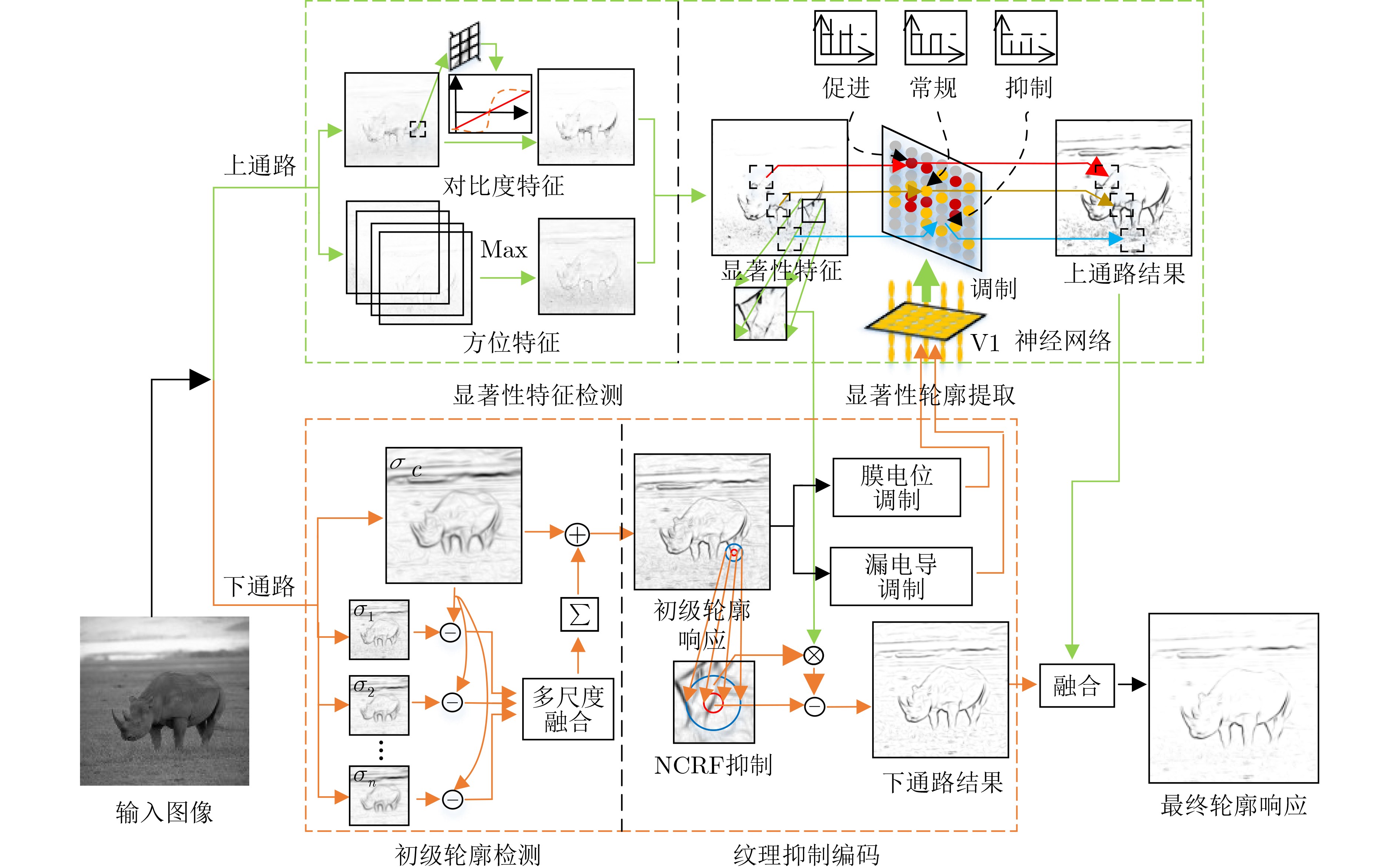
摘要: 基于生物视觉系统存在双视通路(VP)交互感知的机制,该文提出一种图像轮廓检测的新方法。首先针对皮层下视通路中视觉刺激流经多级不同尺度的感受野,提出一种多尺度轮廓融合的轮廓感知模型;接着基于皮层上视通路的对比度适应机制和方向敏感特性,获取显著性视觉特征;然后模拟双视通路的交互感知机制,分别在V1皮层中,构建一种信息流交互引导的脉冲编码模型,提取显著性轮廓;在上丘(SC)浅层提出一种特征调制的非经典感受野侧抑制模型,实现纹理抑制;最后对双视通路中的轮廓响应结果进行修正融合,得到最终轮廓响应。针对RUG40图像库的测试,整个数据集的最优平均P指标和每张图的最优平均P指标分别为0.51和 0.57;针对BSDS500图像库的测试,数据集尺度上最优(ODS)为0.68。结果表明该文方法能有效突显主体轮廓并且抑制纹理背景。通过该文提出的轮廓感知方法,为后续基于视觉机制的图像理解和分析提供了一种新的思路。Abstract: According to the mechanism of visual information interactive perception between dual Visual Pathways(VP) in the biological vision system, a new method of contour detection is proposed. Considering the visual stimulus in the hypocritical pathway flowing through multi-level and different-scale receptive fields, a multi-scale contour fusion contour perception model is proposed. Based on the contrast adaptation mechanism and directional sensitivity of the visual pathway on the cortex, salient visual features are extracted. The interactive perception mechanism of the dual vision pathway is simulated, a pulse coding model is constructed guided by the information flow interaction in the V1 cortex, to extract the saliency contour. An inhibition model of feature modulation non-classical receptive field is proposed in the Superior Colliculus(SC) shallow layer, to achieve texture inhibition. Finally, the contour response results in the dual-view path is modified and fused to obtain the final contour response. For the test of the RUG40 image library, the optimal average P index of the whole dataset and each graph is 0.51 and 0.57 respectively. For the test of the BSDS500 image library, the Optimal Scale (ODS) of the Dataset is 0.68. The results show that the method in this paper can effectively highlight the outline of the subject and suppress the textured background, which provides a new idea for the subsequent image understanding and analysis based on the visual mechanism.
-
Key words:
- Contour detection /
- Interactive perception /
- Contrast adaptive /
- Visual Pathway(VP)
-
表 1 图3中不同算法的参数设置与性能评价指标
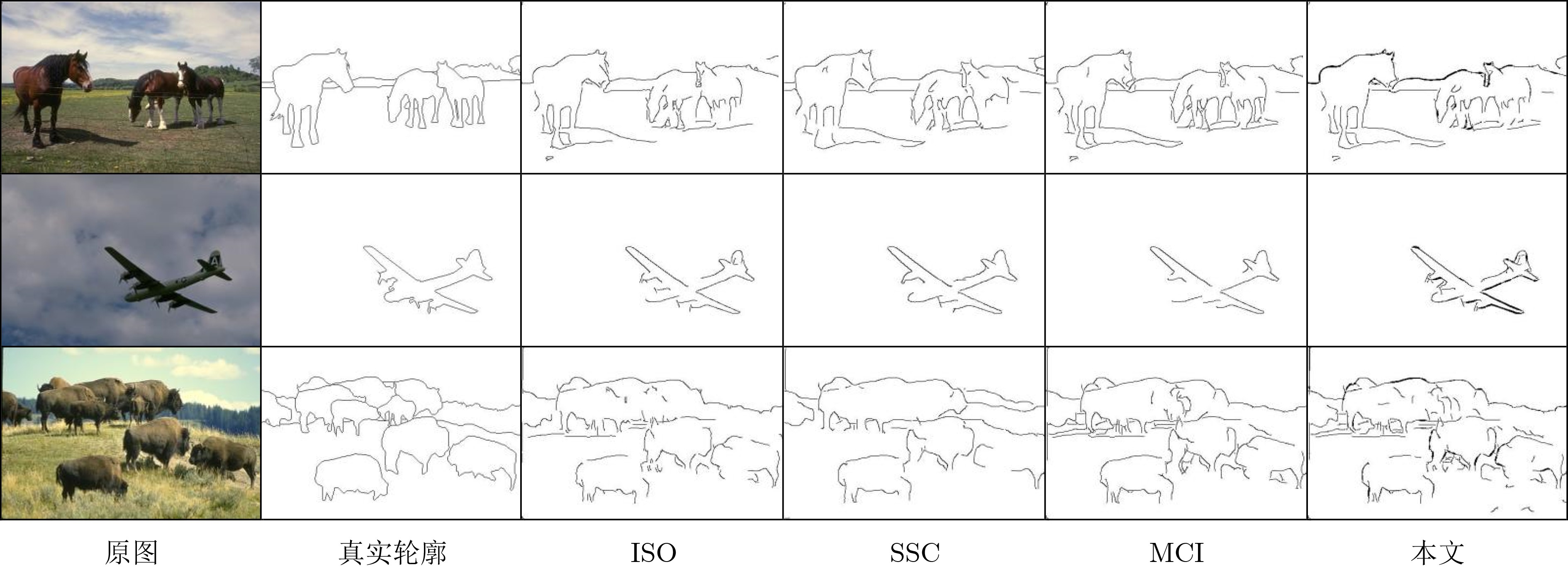
图像 方法 $\alpha $ t ${e_{{\text{FP}}}}$ ${e_{{\text{FN}}}}$ P FPS Buffalo ISO 0.6 0.1 0.25 0.28 0.59 3 MCI 0.7 0.3 0.18 0.29 0.59 1/22 HDC 0.2 0.3 0.16 0.27 0.66 1/27 SNC 0.9 0.2 0.43 0.21 0.58 1/10 本文 0.9 0.6 0.12 0.31 0.69 1/13 Elephant2 ISO 1.0 0.1 0.23 0.36 0.54 MCI 0.4 0.3 0.28 0.33 0.58 HDC 0.2 0.3 0.24 0.29 0.63 SNC 0.9 0.1 0.52 0.24 0.54 本文 1.0 1.0 0.24 0.28 0.64 Hyena ISO 0.9 0.1 0.21 0.27 0.61 MCI 0.6 0.5 0.19 0.22 0.65 HDC 0.1 0.2 0.28 0.20 0.67 SNC 0.9 0.1 0.27 0.22 0.63 本文 0.3 0.5 0.18 0.27 0.71  下载: 导出CSV
-
[1] EDRESS I, AL-SABAWI E A, and YOUNUS M D. Design of fractional-order sobel filters for edge detections[J]. IOP Conference Series:Materials Science and Engineering, 2021, 1152(1): 012028. doi: 10.1088/1757-899X/1152/1/012028 [2] GRIGORESCU C, PETKOV N, and WESTENBERG M A. Contour detection based on nonclassical receptive field inhibition[J]. IEEE Transactions on Image Processing, 2003, 12(7): 729–739. doi: 10.1109/TIP.2003.814250 [3] YANG Kaifu, LI Chaoyi, and LI Yongjie. Multifeature-based surround inhibition improves contour detection in natural images[J]. IEEE Transactions on Image Processing, 2014, 23(12): 5020–5032. doi: 10.1109/TIP.2014.2361210 [4] MELOTTI D, HEIMBACH K, RODRÍGUEZ-SÁNCHEZ A, et al. A robust contour detection operator with combined push-pull inhibition and surround suppression[J]. Information Sciences, 2020, 524: 229–240. doi: 10.1016/j.ins.2020.03.026 [5] CHEN Zekun and CAI Rongtai. Contour detection by simulating the curvature cell in the visual cortex and its application to object classification[J]. IEEE Access, 2020, 8: 74472–74484. doi: 10.1109/ACCESS.2020.2988496 [6] CHEN Shanshan and CAI Houde. Pulvinar involves in multiple pathways of emotion processing[J]. Advances in Psychological Science, 2015, 23(2): 234–240. doi: 10.3724/SP.J.1042.2015.00234 [7] LIDDELL B J, BROWN K J, KEMP A H, et al. A direct brainstem-amygdala-cortical 'alarm' system for subliminal signals of fear[J]. NeuroImage, 2005, 24(1): 235–243. doi: 10.1016/j.neuroimage.2004.08.016 [8] DICARLO J J, ZOCCOLAN D, and RUST N C. How does the brain solve visual object recognition?[J]. Neuron, 2012, 73(3): 425–434. doi: 10.1016/j.neuron.2012.01.010 [9] EVANS H M. The Emotional brain: The mysterious underpinnings of emotional life[J]. Neuropsychoanalysis, 2000, 2(1): 91–95. doi: 10.1080/15294145.2000.10773288 [10] WASSLE H and BOYCOTT B B. Functional architecture of the mammalian retina[J]. Physiological Reviews, 1991, 71(2): 447–480. doi: 10.1152/physrev.1991.71.2.447 [11] WANG Luping, SARNAIK R, RANGARAJAN K, et al. Visual receptive field properties of neurons in the superficial superior colliculus of the mouse[J]. Journal of Neuroscience, 2010, 30(49): 16573–16584. doi: 10.1523/JNEUROSCI.3305-10.2010 [12] DE FRANCESCHI G and SOLOMON S G. Visual response properties of neurons in the superficial layers of the superior colliculus of awake mouse[J]. The Journal of Physiology, 2018, 596(24): 6307–6332. doi: 10.1113/JP276964 [13] LUSSIEZ R, CHANAURIA N, OUELHAZI A, et al. Effects of visual adaptation on orientation selectivity in cat secondary visual cortex[J]. The European Journal of Neuroscience, 2021, 53(2): 588–600. doi: 10.1111/ejn.14967 [14] AHMADLOU M, ZWEIFEL L S, and HEIMEL J A. Functional modulation of primary visual cortex by the superior colliculus in the mouse[J]. Nature Communications, 2018, 9(1): 3895. doi: 10.1038/s41467-018-06389-6 [15] HAN J K, KIM M S, KIM S I, et al. Investigation of leaky characteristic in a single-transistor-based leaky integrate-and-fire neuron[J]. IEEE Transactions on Electron Devices, 2021, 68(11): 5912–5915. doi: 10.1109/TED.2021.3110830 [16] ALPERT S, GALUN M, BRANDT A, et al. Image segmentation by probabilistic bottom-up aggregation and cue integration[J]. IEEE Transactions on Pattern Analysis and Machine Intelligence, 2012, 34(2): 315–327. doi: 10.1109/TPAMI.2011.130 [17] KANEDA K, YANAGAWA Y, and ISA T. Inputs from visual cortex contribute to lateral inhibition in the mouse superior colliculus in vivo[J]. Neuroscience Research, 2009, 65(S1): S172. doi: 10.1016/j.neures.2009.09.905 [18] CHEN Yanmei, NI Yiling, ZHOU Jianhong, et al. The amygdala responds rapidly to flashes linked to direct retinal innervation: A flash-evoked potential study across cortical and subcortical visual pathways[J]. Neuroscience Bulletin, 2021, 37(8): 1107–1118. doi: 10.1007/s12264-021-00699-4 [19] CHEN Shu'nan, FAN Yingle, FANG Tao, et al. A contour detection method based on hierarchical structure response model in primary visual pathway[J]. Acta Automatica Sinica, 2022, 48(3): 820–833. doi: 10.16383/j.aas.c200046 [20] MARTIN D R, FOWLKES C C, and MALIK J. Learning to detect natural image boundaries using local brightness, color, and texture cues[J]. IEEE Transactions on Pattern Analysis and Machine Intelligence, 2004, 26(5): 530–549. doi: 10.1109/TPAMI.2004.1273918 [21] LIU Yun, CHENG Mingming, HU Xiaowei, et al. Richer convolutional features for edge detection[J]. IEEE Transactions on Pattern Analysis and Machine Intelligence, 2019, 41(8): 1939–1946. doi: 10.1109/TPAMI.2018.2878849 [22] XIE Saining and TU Zhuowen. Holistically-nested edge detection[C]. 2015 IEEE International Conference on Computer Vision (ICCV), Santiago, Chile, 2015: 1395–1403. [23] YANG Kaifu, GAO Shaobing, GUO Cefeng, et al. Boundary detection using double-opponency and spatial sparseness constraint[J]. IEEE Transactions on Image Processing, 2015, 24(8): 2565–2578. doi: 10.1109/TIP.2015.2425538 -

 下载:
下载:


图(6) / 表(1)
计量
- 文章访问数: 376
- HTML全文浏览量: 282
- PDF下载量: 52
- 被引次数: 0
